. 2010 Apr 16;141(2):268-79.
doi: 10.1016/j.cell.2010.03.021.

Periodic cyclin-Cdk activity entrains an autonomous Cdc14 release oscillator

Affiliations

Periodic cyclin-Cdk activity entrains an autonomous Cdc14 release oscillator

Ying Lu et al. Cell. .

Abstract

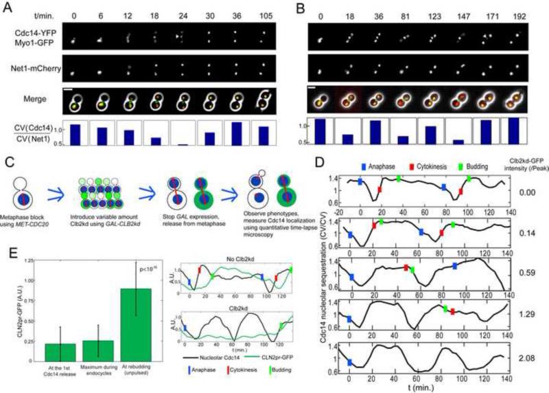
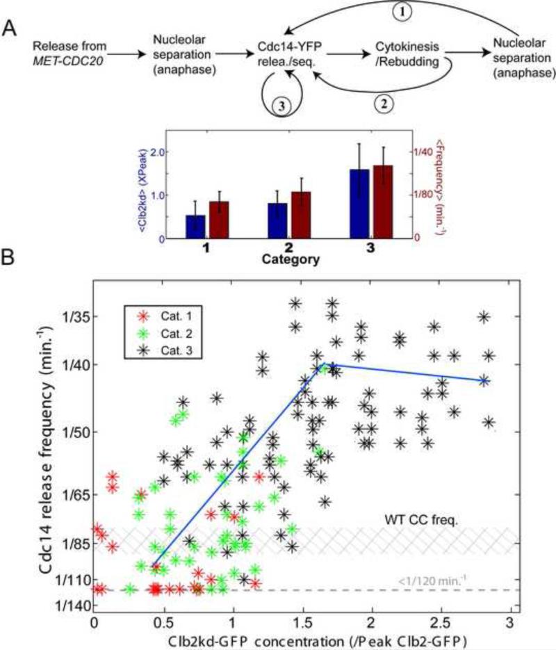
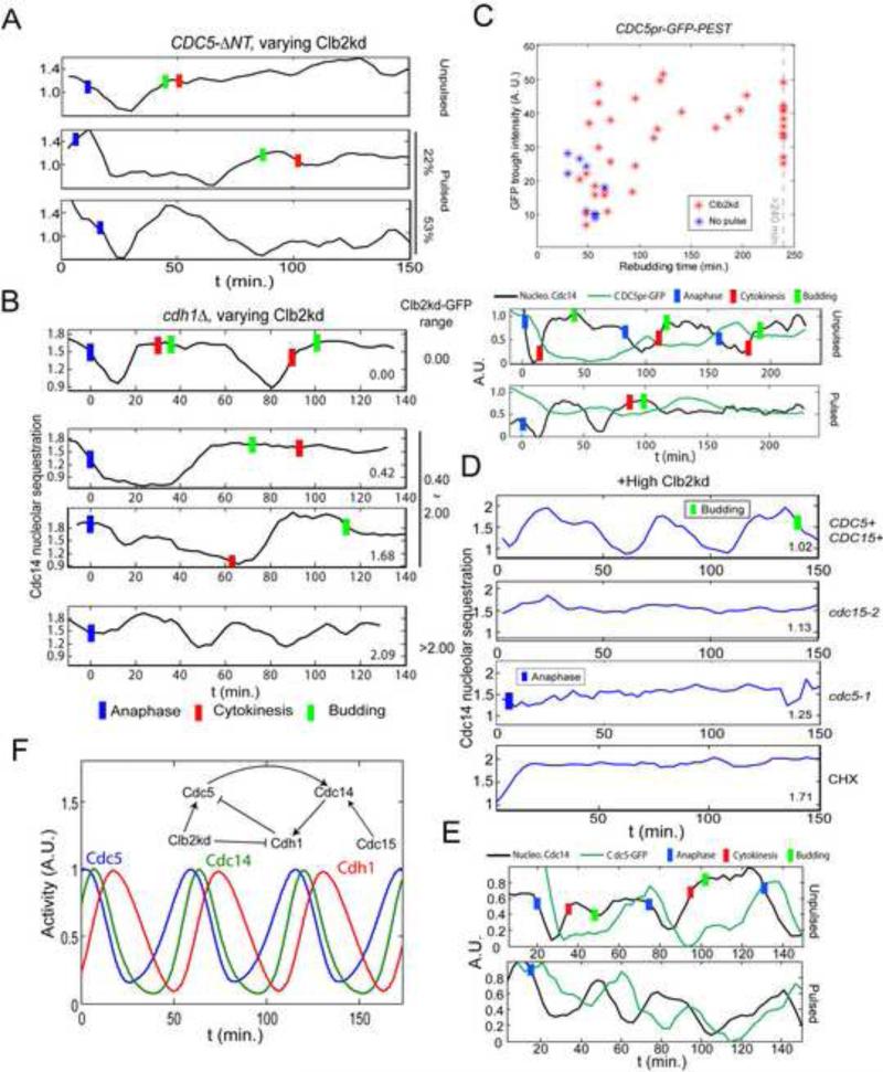
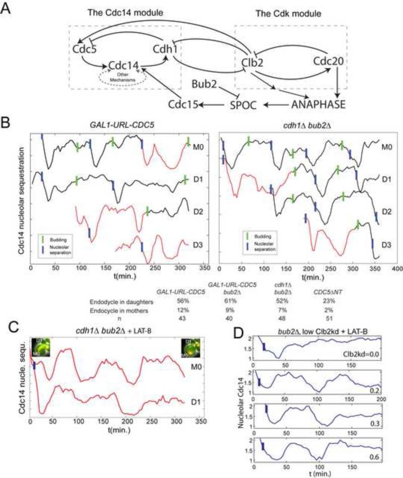
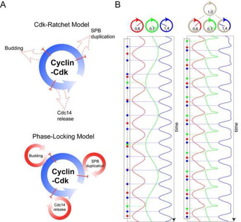
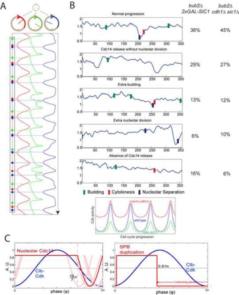
One oscillation of Cyclin-dependent kinase (Cdk) activity, largely driven by periodic synthesis and destruction of cyclins, is tightly coupled to a single complete eukaryotic cell division cycle. Tight linkage of different steps in diverse cell-cycle processes to Cdk activity has been proposed to explain this coupling. Here, we demonstrate an intrinsically oscillatory module controlling nucleolar release and resequestration of the Cdc14 phosphatase, which is essential for mitotic exit in budding yeast. We find that this Cdc14 release oscillator functions at constant and physiological cyclin-Cdk levels, and is therefore independent of the Cdk oscillator. However, the frequency of the release oscillator is regulated by cyclin-Cdk activity. This observation together with its mechanism suggests that the intrinsically autonomous Cdc14 release cycles are locked at once-per-cell-cycle through entrainment by the Cdk oscillator in wild-type cells. This concept may have broad implications for the structure and evolution of eukaryotic cell-cycle control.


Figures

Figure 1
Figure 1. Cyclical Cdc14 release uncoupled from cell cycle progression
A, B. MET3-CDC20 CDC14-YFP NET1-mCherry MYO1-GFP cells were released from a MET3-CDC20 block (t=0). Bottom: Cdc14 release was quantified at each time point as the following: the coefficient of variation (CV) of Cdc14-YFP signal inside a single cell, computed from fluorescent time-lapse microscopy data, is the standard deviation of YFP pixel intensity across the cell, divided by the mean intensity; CV of Cdc14-YFP is then divided by CV of Net1-mCherry, and this ratio will be high in cells with Cdc14-YFP concentrated in specific regions, and low when Cdc14-YFP is dispersed through the cell. Triangle: disappearing Myo1 ring. Scale bar: 5 microns. A: control. B. Clb2kd was pulsed for 30 min. before release. C. Schematic of procedure for loading cells with undegradable Clb2kd (green) before ME. Nucleus is shown in blue, spindle in red D. Pulsed Clb2kd-GFP was quantified (right column) in units standardized to the peak level of Clb2 attained in a normal cell cycle, and Cdc14 localization quantified (n=170). Blue bars: anaphase (nucleolar separation, marked by Net1-mCherry); Red bars: cytokinesis (Myo1 ring disappearance); Green bars: bud emergence. E. CLN2 promoter expression during Cdc14 endocycles. A CLN2pr-GFP-PEST strain was pulsed with Clb2kd as in (B) for 35 minutes. GFP intensities at the first Cdc14 release, maximum during endocycles (n=40), and at rebudding in unpulsed control cells (P<10−15. Error bars: standard deviation). See also figure S1 and movie S1.
Figure 2
Figure 2. Clb2kd level controls the Cdc14 endocycle period
A. Trajectories for Clb2kd-pulsed cells. Category 1: essentially normal cell-cycle progression and Cdc14 release/sequestration; Category 2: a second Cdc14 release occurred between rebudding and nucleolar separation in the next cell cycle; Category 3: Cdc14 endocycles without cytokinesis or rebudding. Below: category means and standard deviations of Clb2kd-GFP concentration (blue) and Cdc14 release frequencies (red). B. Cdc14 release frequencies plotted against Clb2kd-GFP level for cell categories: inverse of intervals between first and second Cdc14 release (categories 1 and 2), or average frequencies of one cell's Cdc14 endocycles (category 3; correlation between the1st and 2nd cycle is shown in Fig. S2B). Shaded: range of cell cycle frequencies for cycling MET3-CDC20 mother cells. 31/170 category 1; 45/170 category 2; 94/170 category 3.. See also figure S2.
Figure 3
Figure 3. Requirements for Cdc14 endocycles
A. As in Fig. 1B, but cells also cdc5::3XCDC5-ΔNT. Among cells whose rebudding was delayed for at least 25 minutes (implying >1X peak Clb2kd, Fig. S1B), 19/36 cells showed fast Cdc14 release and resequestration, followed by prolonged Cdc14 release. 8/36 showed only prolonged Cdc14 release. B. As in Fig. 1D, but also cdh1Δ; 30 min Clb2kd-GFP pulse; typical traces for the indicated Clb2kd-GFP ranges. Among cells with >1X peak Clb2kd-GFP (n=86), 41% showed a prolonged Cdc14 release period (middle two traces); 43% (cells with highest Clb2kd-GFP) showed release endocycles with a reduced amplitude (bottom trace); Blue bars: anaphase; Red bars: cytokinesis; Green bars: bud emergence. C. CDC5pr-GFP cells, as in Fig. 1B. Trough GFP intensities before rebudding plotted against rebudding times; rebudding delay indicates Clb2kd levels; sample traces below. D. Cells as in Fig. 1D, but also cdc15-2 or cdc5-1; after 35 min to allow initial Cdc14 release at permissive temperature, cells were plated for time-lapse at 37°C (restrictive temperature) (t=0). CHX: as above, except that time-lapse medium contained 0.2ng/μl cycloheximide (CHX). Among cells with >1X peak Clb2kd-GFP, 18/24 CDC15 CDC5 cells, 0/22 cdc15-2, 0/30 cdc5-1 and 3/64 cells in CHX exhibited Cdc14 release endocycles. Blue bars: anaphase; Green bars: bud emergence. E: As in Fig. 1B, but CDC5-GFP cells; 30 min Clb2kd pulsed; typical traces of Cdc14 release and Cdc5-GFP levels are shown. In 36/45 Cdc14-endocycling cells, Cdc5-GFP signal oscillated out-of-phase with Cdc14 release. F. ODE model simulating Cdc14-Cdh1-Cdc5 negative feedback (Supplemental Methods). See also figure S3.
Figure 4
Figure 4. Cdc14 release endocycles in cycling cells or with low Clb2kd
A. Cdc14 release and Clb-Cdk control mechanisms. Left: potential autonomous Cdc14 release oscillator; right: Clb-Cdk negative-feedback oscillator. SPOC: spindle orientation checkpoint. B. Cdc14 release analyzed as in Fig. 1, in bub2Δ cdh1Δ and GAL1-URL-CDC5 cells (cycling cells, without cdc20 block-release or Clb2kd pulse). A representative lineage (M0: mother, D1, D2...sequential daughters) exhibiting ectopic Cdc14 release endocycles (red) before bud emergence. Cdc14 release curves shifted for visualization. Below: probabilities of G1 Cdc14 endocycles. C. 67μM Latrunculin-B (LAT-B) was added to the medium (t=0) to inhibit budding of a cycling bub2Δ cdh1Δ strain. 32/44 cells demonstrated G1 Cdc14 endocycles. 14/19 daughter cells and 13/19 mother cells exhibited endocycles (maximum 4 endocycles; average=2.8). Cell images at the beginning/end of the time-course indicates no cell cycle progression. D. MET-CDC20 bub2Δ cdc15-2 cells were arrested in metaphase at 34°C and pulsed with Clb2kd-GFP for 20 minutes (using cdc15-2 to achieve a stable block), then released into cell cycle at 28°C with 67μM LAT-B. First anaphase (nucleolar separation) happened with normal kinetics. LAT-B effectively blocked budding, cytokinesis, and subsequent anaphase in ~90% cells. 39/46 cells containing 0.2~1.0x peak Clb2kd-GFP demonstrated Cdc14 endocycles. Four traces are shown. Blue bars: anaphase. See also figure S4.
Figure 5
Figure 5. Cell cycle control through phase-locking
A. Schematic of ratchet (above) and phase-locking (below). B. Conceptual model: three simply oscillators ((Kuramoto, 1975), Supplemental Methods) with different intrinsic frequencies (indicated) control different cell cycle events. Assuming a single event is generated as the oscillator's phase evolves passing n*2π or ‘12 o’clock’. Without entrainment by cyclin-Cdk oscillator, each peripheral oscillator evolves uniformly, and there is no fixed order (left) among events they generate. Allowing a cyclin-Cdk oscillator to advance or delay part of the peripheral oscillators’ cycles leads to phase-locking and a stable order of events (Right). See also figure S5.
Figure 6
Figure 6. Experimental test of phase-locking predictions
A. Model as in Fig. 5B, but the amplitude of cyclin-Cdk oscillation is reduced by 15%, resulting in event disorder (compare Fig. 5B, right). B. 2XGAL1-SIC1 bub2Δ or cdh1Δ sic1Δ bub2Δ strains may reduce the amplitude of cyclin-Cdk oscillation by lowering the peak Cdk activity or raising the trough (cartoon below). 2XGAL1-SIC1 cells (n=37) were grown in raffinose medium prior to time-lapse analysis on galactose medium to induce Sic1 (t=0). cdh1Δ sic1Δ GALL-CDC20 cells (n=35) were imaged on glucose to turn off GALL-CDC20. Cells were classified according the cell cycle disorder that appeared first, shown with representative traces (right). Blue bars: nucleolar separation; Green bars: budding; Red bars: cytokinesis. C. Cdc14 release and SPB duplication timing in free-running cell cycles predicted by a quantitative phase-locking model. Red: best estimation of parameters; blue: sine wave simulation of Clb-Cdk cycle. Light-red: solutions with parameters altered +/− one standard deviation for one or both of the two parameters (for two combinations no solution could be obtained due to mathematical restrictions of the model; Supplemental Methods). Right panel: phase-locking could entrain SPB duplication oscillator (red) to the correct position early in the Clb-Cdk cycle (blue) (Supplemental Methods). See also figure S6.

Comment in

Similar articles

Cited by

References

    1. Alexandru G, Zachariae W, Schleiffer A, Nasmyth K. Sister chromatid separation and chromosome re-duplication are regulated by different mechanisms in response to spindle damage. The EMBO journal. 1999;18:2707–2721. - PMC - PubMed
    1. Azzam R, Chen SL, Shou W, Mah AS, Alexandru G, Nasmyth K, Annan RS, Carr SA, Deshaies RJ. Phosphorylation by cyclin B-Cdk underlies release of mitotic exit activator Cdc14 from the nucleolus. Science (New York, NY. 2004;305:516–519. - PubMed
    1. Bardin AJ, Visintin R, Amon A. A mechanism for coupling exit from mitosis to partitioning of the nucleus. Cell. 2000;102:21–31. - PubMed
    1. Bean JM, Siggia ED, Cross FR. Coherence and timing of cell cycle start examined at single-cell resolution. Molecular cell. 2006;21:3–14. - PubMed
    1. Bhattacharyya RP, Remenyi A, Yeh BJ, Lim WA. Domains, motifs, and scaffolds: the role of modular interactions in the evolution and wiring of cell signaling circuits. Annual review of biochemistry. 2006;75:655–680. - PubMed

Publication types

MeSH terms