. 2010 Apr 16;141(2):304-14.
doi: 10.1016/j.cell.2010.02.035.

LIS1 and NudE induce a persistent dynein force-producing state

Affiliations

LIS1 and NudE induce a persistent dynein force-producing state

Richard J McKenney et al. Cell. .

Abstract

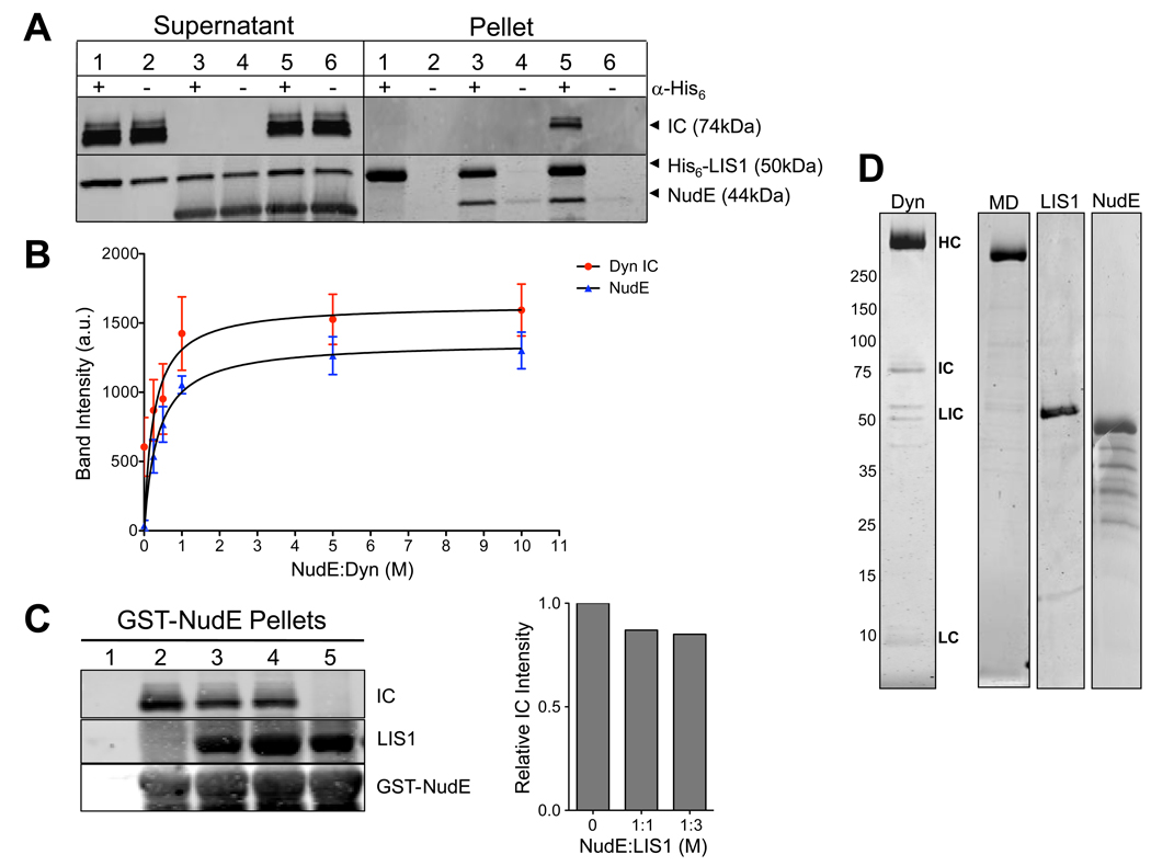
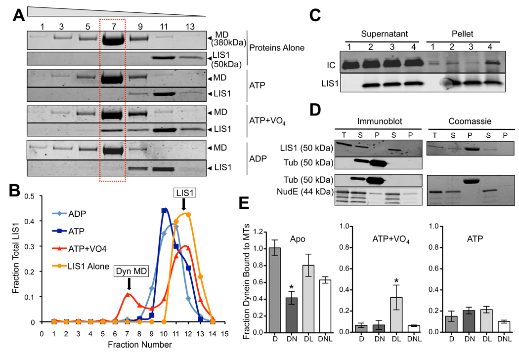
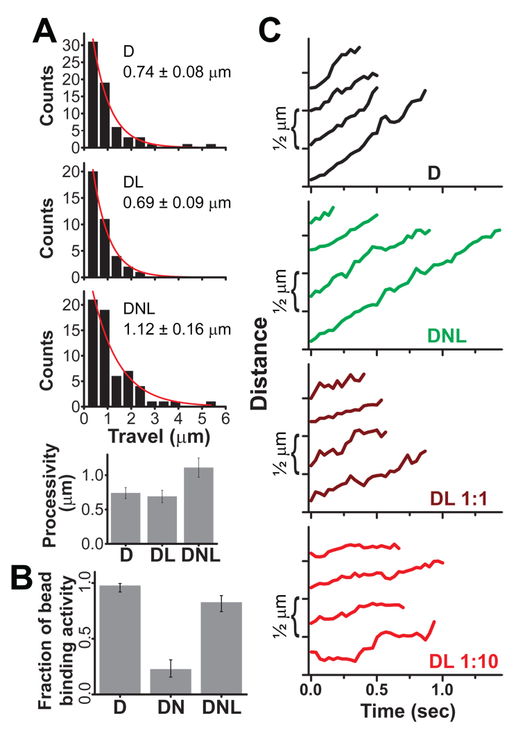
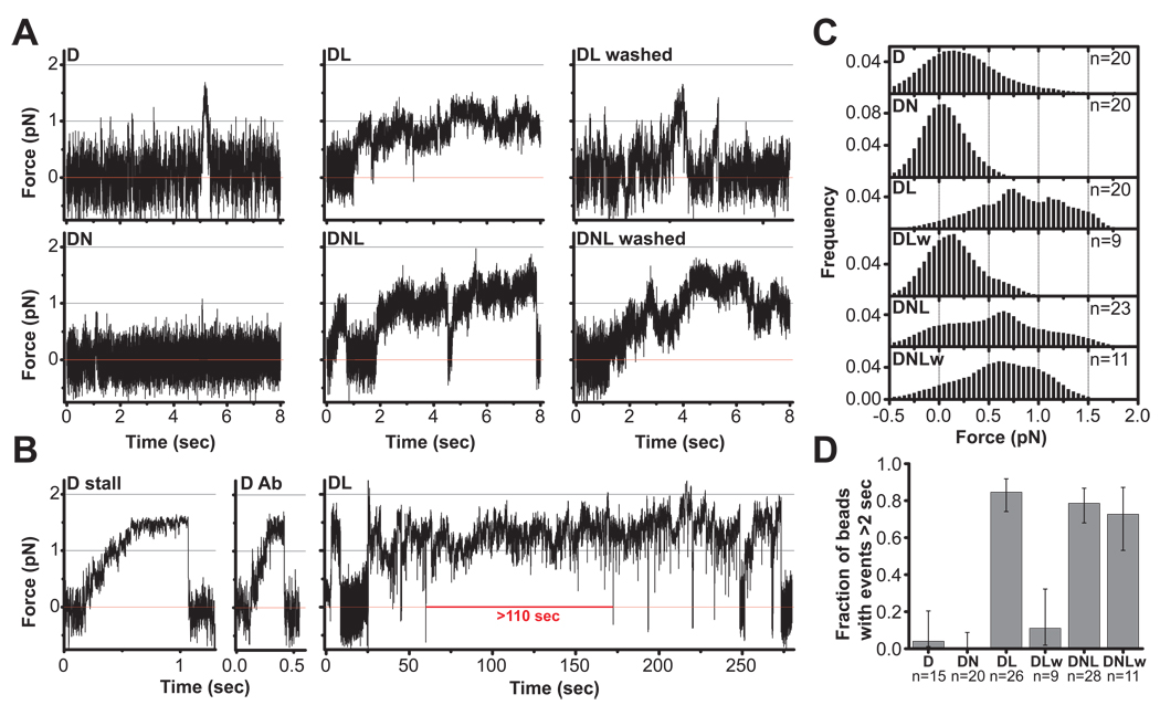
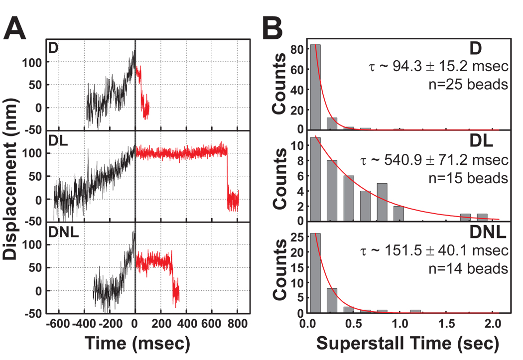
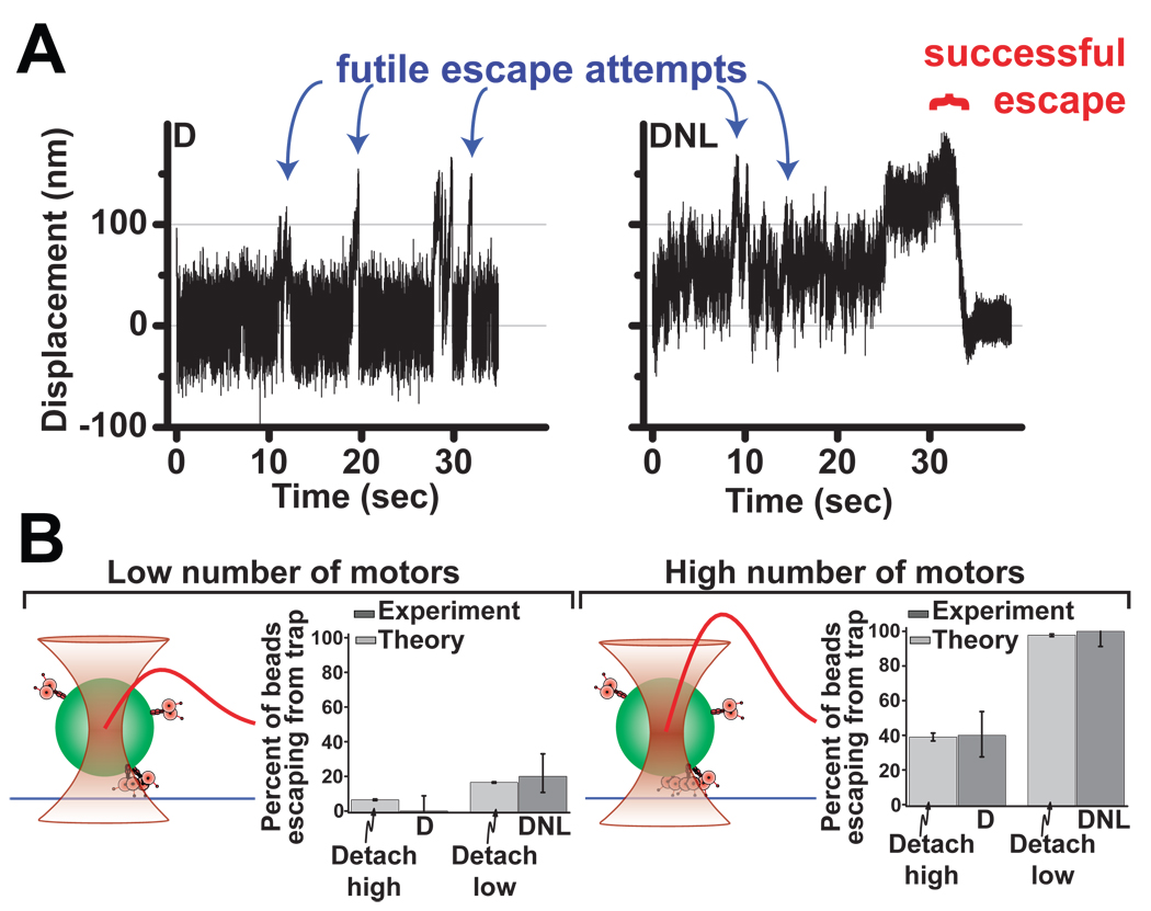
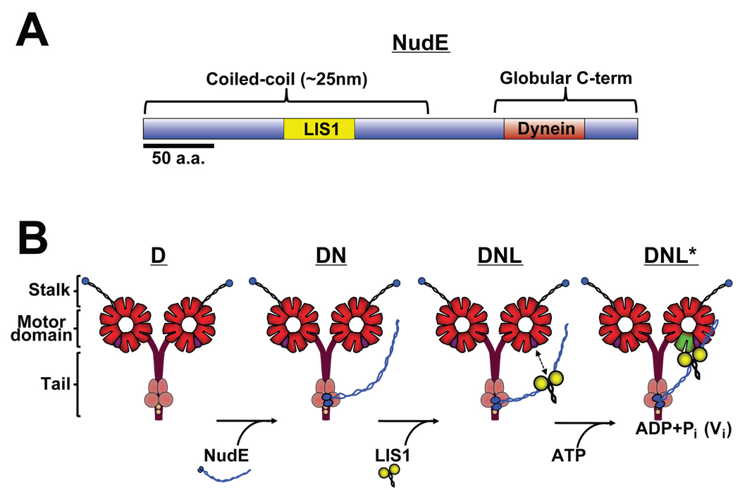
Cytoplasmic dynein is responsible for many aspects of cellular and subcellular movement. LIS1, NudE, and NudEL are dynein interactors initially implicated in brain developmental disease but now known to be required in cell migration, nuclear, centrosomal, and microtubule transport, mitosis, and growth cone motility. Identification of a specific role for these proteins in cytoplasmic dynein motor regulation has remained elusive. We find that NudE stably recruits LIS1 to the dynein holoenzyme molecule, where LIS1 interacts with the motor domain during the prepowerstroke state of the dynein crossbridge cycle. NudE abrogates dynein force production, whereas LIS1 alone or with NudE induces a persistent-force dynein state that improves ensemble function of multiple dyneins for transport under high-load conditions. These results likely explain the requirement for LIS1 and NudE in the transport of nuclei, centrosomes, chromosomes, and the microtubule cytoskeleton as well as the particular sensitivity of migrating neurons to reduced LIS1 expression.


Figures

Fig. 1
Fig. 1. Dynein-NudE-LIS1 interactions
(A) Recruitment of LIS1 to dynein by NudE. His6-tagged LIS1 was immunoprecipitated with anti-His6 antibody in the presence of purified brain dynein, NudE, or both. Immunoblotting shows no detectable coimmunoprecipitation of dynein (visualized by anti-intermediate chain (IC) antibody) with LIS1 (Pellet, lane 1) unless NudE is included (Pellet, lane 5). (B) NudE concentration dependence of LIS1-dynein interaction. NudE was added in increasing amounts to a mixture of LIS1 and dynein, and the mixture was adsorbed to protein A beads coated with LIS1 antibodies. Quantitation of dynein (IC) and NudE pulled-down with LIS1 is shown. Dynein binding saturated at ~1:1 molar ratio with NudE. Mean of raw band intensity from 4 experiments is plotted in arbitrary units (a.u.) +/− SD. (C) LIS1 and dynein do not compete for NudE. GST-NudE-coated beads were mixed with dynein either in the absence of LIS1 (lane 2) or the presence of 1 (lane 3) or 3-fold (lane 4) molar stoichiometry of LIS1:NudE. The added LIS1 did not substantially affect the amount of dynein that bound to NudE confirming that NudE can bind both LIS1 and dynein simultaneously (see Fig. 7A). No proteins bound to beads without NudE (lane 1) and LIS1 could bind to NudE in the absence of dynein (lane 5). Right, quantification of the relative amount of dynein IC in the absence and presence of added LIS1 protein. (D) Coomassie brilliant blue stained gels of purified cytoplasmic dynein complex and recombinant proteins used in this study. MD: Purified dynein motor domain. Dynein subunits: HC - heavy chain; IC - intermediate chain; LIC - light intermediate chain; LC – light chain. Bands below NudE represent fragments, as judged by immunoblotting with anti-NudE antibody. See also Fig. S1.
Fig. 2
Fig. 2. Effect of Nucleotides on Dynein-NudE-LIS1 interactions
(A) Effect of nucleotides on LIS1 binding to dynein motor domain. Purified baculovirus-expressed dynein motor domain (MD) and LIS1 were incubated separately or together and sedimented through sucrose gradients containing the indicated nucleotides. Coomassie brilliant blue stained gels show a fraction of LIS1 to co-sediment with the purified dynein motor domain peak (dashed box) in the presence of ATP and VO4, but not ATP alone or ADP. Fraction numbers are indicated at top. (B) Quantitation of LIS1 distribution showing a major low s-value peak for the free protein, and, in the ATP+VO4 condition, a smaller peak cosedimenting with the dynein motor domain. (C) Effect of nucleotides on LIS1 binding to purified calf brain dynein. Dynein was incubated with beads alone (lane 1) or LIS1-coated beads in the absence of nucleotide (lane 2) or in the presence of ATP (lane 3) or ATP+VO4 (lane 4). Dynein (IC) is enriched in the LIS1 pellet only in the ATP+VO4 condition. (D) LIS1 and NudE do not bind to microtubules. Purified LIS1 and NudE were sedimented in the presence or absence of microtubules. Total protein (T), supernatants (S) and pellets (P) are shown by immunoblotting and Coomassie blue staining. LIS1 is obscured by tubulin in the Coomassie blue-stained gel, but immunoblotting shows LIS1 not to sediment with microtubules. NudE also shows no evidence of microtubule cosedimentation. (E) Effect of LIS1 and NudE on dynein binding to microtubules. Purified brain dynein was mixed with microtubules in the absence of nucleotide (Apo) or in the presence of ATP or ATP+VO4, and the microtubules were sedimented. NudE strongly inhibited binding of dynein to microtubules in the apo state (DN; *P = 0.0011 two-tailed t-test), an effect partially rescued by LIS1 (DNL). Conversely, LIS1 increased dynein binding to microtubules in the ATP+VO4 state by approximately 5-fold (DL; * P = 0.0131 two-tailed t-test). Error bars represent mean +/− SD of 3 experiments.
Fig. 3
Fig. 3. Effect of LIS1 and NudE on Dynein Motility
(A) Histogram of single dynein travel distances alone (D), in the presence of LIS1 (molar ratio of D:L = 1:2), and in the presence of both NudE and LIS1 (D:N:L = 1:9:10). Exponential decay fits are shown in red, and decay constant ± SEM is indicated in each case. A small percentage of beads had bidirectional motion of 2 µm or more in each direction, did not spontaneously detach from microtubules, and were excluded from analysis; such beads showed no force production capacity in either direction and were deemed to be diffusive rather than processive. D and DL processivity values are similar, whereas DNL processivity is clearly increased (P <0.03). (B) Microtubule-binding activity for D, DN, and DNL beads. We quantify the percentage of beads with visible binding and travel events, held close to a microtubule in a weak optical trap (See Methods). NudE (molar ratio 1:9 D:N) potently reduced microtubule binding activity, an effect rescued by addition of LIS1 (1:9:2 D:N:L). Neither LIS1 nor NudE produced a comparable inhibition of binding activity in similar experiments with kinesin motors (Fig. S2B). Exact CI error bars are reported (see Supplement). (C) Individual traces of motor-driven movements along microtubules (D:L ratios indicated; D:N:L is as in A). D (black) and DNL (green) beads show robust motility whereas DL bead movements (dark and light red) are interrupted by pauses that result in a net slow-down of transport. The effect becomes more prominent as the LIS1 concentration increases. See Fig. S3 for more extended and extreme examples of bead travel. For all experiments, dynein was first adsorbed to beads, followed as needed by NudE and then by LIS1.
Fig. 4
Fig. 4. The effect of NudE and LIS1 on dynein force production
(A) Representative records of single motor force production in an optical trap for beads with bound dynein (D); dynein + NudE (DN); dynein + LIS1 (DL); and dynein + NudE + LIS1 (DNL). Red line marks center of optical trap. Dynein beads (D) typically detach before the motor can stall, but short (~0.5 sec) stalls occasionally occur (see B). DN beads (1:9 D:N) show almost no motion. However, DL and DNL beads (1:2 D:L and 1:9:2 D:N:L) exhibited dramatically longer force production events, some of which continued beyond the period shown (and see panel B). Similar behavior under load was also observed at higher amounts of LIS1 (1:10 D:L) in DL and DNL assays (see Fig. S4). The prolonged dynein stalls induced by LIS1 were eliminated by washing the beads (see Methods) in the DL assay (DL washed) but not the DNL assay (DNL washed). These observations support our biochemical results indicating that NudE retains LIS1 in a complex with dynein (Fig. 1A, B). (B) Representative stalls for dynein adsorbed to beads nonspecifically (D stall) or through anti-dynein IC monoclonal antibody (D Ab) illustrate maximal force production for single dynein motors. (DL) An extremely long (~110 sec) event (entire tracing is shown) demonstrating dramatic prolongation of dynein force production by LIS1. (C) Distribution of forces attained during prominent bead-microtubule binding events sampled in a 4 second window, and summed from multiple bead assays (n= 9–23 as indicated). DN shows minimal bead displacement almost symmetrically distributed around the trap center, indicating that bead motion is predominantly due to thermal noise. D alone exhibits a shift to higher forces. The shift is dramatically greater for DL and DNL, indicating higher average force production. Notably, this effect was retained when DNL beads were washed to remove excess proteins from solution (DNLw) but abolished when DL beads were similarly washed (DLw). This suggests that DNL association is stable but the DL one is not. (D) The fraction of beads exhibiting long force production events (>2 sec) is dramatically increased in the presence of LIS1. The effect is again abolished in DL-washed (DLw) but not in DNL-washed (DNLw) assays. Exact CI error bars are reported (see Supplement). See also Figure S4.
Fig. 5
Fig. 5. LIS1 enhances dynein-microtubule interaction under load
(A) Beads driven by single dynein motors were allowed to move along microtubules in a weak optical trap (black traces). At 100 nm bead displacement from the trap center, laser power was automatically increased (T=0, solid vertical line), subjecting dynein to a load of ~2 pN, significantly greater than the dynein stall force (“superstall” conditions). Subsequent bead positions are shown as red lines, and demonstrate prolonged persistence of DL bead (1:10 D:L) on microtubule and less so of DNL (1:9:10 D:N:L) bead. (B) Analysis of multiple superstall traces as in (A) revealed that average detachment times in both DL and DNL assays were significantly increased relative to D alone. Exponential decay constant ± SEM is shown in each subpanel.
Fig. 6
Fig. 6. LIS1 and NudE enhance multiple motor function
To test the effect of NudE and LIS1 on multiple dynein motors, beads were incubated with concentrations of dynein above those used for single molecule experiments. (A) Force records from individual beads exposed to dynein (D) or the same amount of dynein followed by NudE and LIS1 (DNL). High force events can be observed (blue arrows), as can bead escapes from trap confinement (red bracket). (B) Quantification of trap escape. The fraction of high force events resulting in escape was scored for D vs. DNL at low (left) and high (right) concentrations of applied dynein (dark gray bars, n=20 in each case). Maximum trap force was estimated at 1.8 pN and 3.7 pN for these two assays respectively (therefore, synergistic activity of ≥2 and ≥3 motors was required for a successful escape event to occur in these two assays, respectively). The frequency of trap escapes was clearly increased in DNL vs. D alone. Theoretical modeling (light gray bars) conservatively assuming a 50% change of time to detachment for each dynein motor (cf Fig. 5B) is predicted to be sufficient to account for the observed difference between D and DNL escape frequencies (see supplementary information). Schematic diagram of the experiment (left of each histogram): the bead (green) with motors attached to its surface (rose) is held near a microtubule (blue) by an optical trap (hyperboloid shape). Trap strength (red curve) rises approximately linearly as the distance from trap center grows, then saturates and finally decays to zero. Exact CI error bars are reported (see Supplement). See also Figure S5.
Fig. 7
Fig. 7. Diagrammatic representation of dynein-LIS1-NudE interactions and functional consequences
(A) Bar diagram of NudE shows coiled-coil and unstructured C-terminal domains, and known LIS1 and dynein binding regions (see text for details). (B) Proposed assembly intermediates in dynein-NudE-LIS1 complex. Known binding of NudE to dynein intermediate and light chains (Stehman et al., 2007) predicts association of NudE C-terminus with base of the dynein molecule and protruding coiled-coil domain as shown. The calculated distance between this site and the known LIS1 binding site in the middle of the NudE coiled-coil α-helical tail (Derewenda et al., 2007) is proposed here to be sufficient to allow NudE to position LIS1 near the dynein motor domains. LIS1 is shown unbound to the dynein motor domain as we observe under most conditions, but is proposed to bind specifically in the ADP-VO4 prepowerstroke state (Fig. 2).

Similar articles

Cited by

References

    1. Bremner KH, Scherer J, Yi J, Vershinin M, Gross SP, Vallee RB. Adenovirus transport via direct interaction of cytoplasmic dynein with the viral capsid hexon subunit. Cell Host Microbe. 2009;6:523–535. - PMC - PubMed
    1. Burgess SA, Walker ML, Sakakibara H, Knight PJ, Oiwa K. Dynein structure and power stroke. Nature. 2003;421:715–718. - PubMed
    1. Chen B, Doucleff M, Wemmer DE, De Carlo S, Huang HH, Nogales E, Hoover TR, Kondrashkina E, Guo L, Nixon BT. ATP ground- and transition states of bacterial enhancer binding AAA+ ATPases support complex formation with their target protein, sigma54. Structure. 2007;15:429–440. - PMC - PubMed
    1. Coquelle FM, Caspi M, Cordelieres FP, Dompierre JP, Dujardin DL, Koifman C, Martin P, Hoogenraad CC, Akhmanova A, Galjart N, et al. LIS1, CLIP-170's key to the dynein/dynactin pathway. Mol Cell Biol. 2002;22:3089–3102. - PMC - PubMed
    1. Derewenda U, Tarricone C, Choi WC, Cooper DR, Lukasik S, Perrina F, Tripathy A, Kim MH, Cafiso DS, Musacchio A, Derewenda ZS. The structure of the coiled-coil domain of Ndel1 and the basis of its interaction with Lis1, the causal protein of Miller-Dieker lissencephaly. Structure. 2007;15:1467–1481. - PubMed

Publication types