. 2010 Jun;120(4-5):172-9.
doi: 10.1016/j.jsbmb.2010.04.009. Epub 2010 Apr 18.

The tri-nucleotide spacer sequence between estrogen response element half-sites is conserved and modulates ERalpha-mediated transcriptional responses

Affiliations

The tri-nucleotide spacer sequence between estrogen response element half-sites is conserved and modulates ERalpha-mediated transcriptional responses

Feng-Jue Shu et al. J Steroid Biochem Mol Biol. 2010 Jun.

Abstract

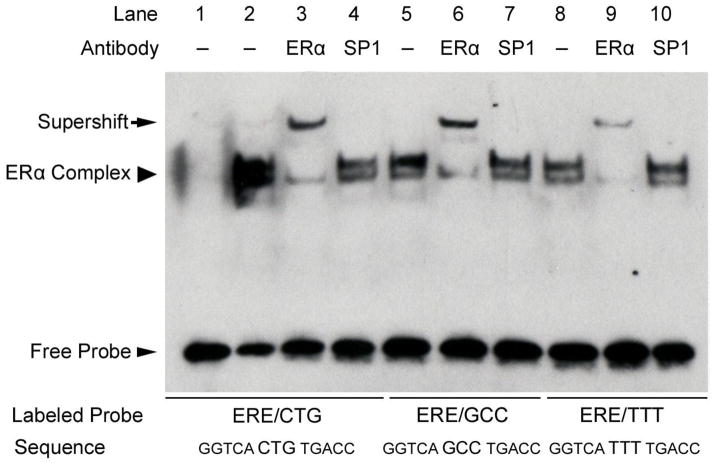
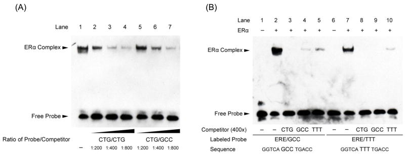
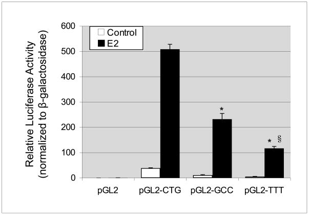
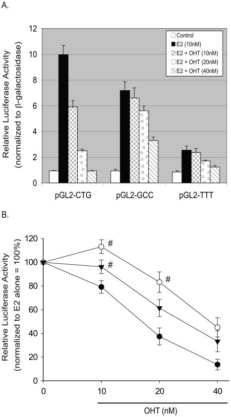
The estrogen response element (ERE) consensus sequence is AGGTCAnnnTGACCT, where nnn is known as the tri-nucleotide spacer sequence. Studying 1017 high-confidence ERalpha-bound loci, we found that genomic EREs are enriched for spacers composed of C(A/T)G, suggesting that the spacer may influence receptor binding and transcriptional responses. We designed consensus EREs containing variable spacer sequences and compared ERalpha binding in gel shift assays and enhancer function in reporter assays. We found that ERalpha-ERE binding affinity is modulated by the tri-nucleotide spacer sequence and is favored by spacer sequences of CTG>GCC>TTT. Similarly, luciferase reporter assays indicated that the estrogen-stimulated transcriptional response is modulated by the spacer and parallels the gel shift data: CTG>GCC>TTT. Reporter assays demonstrated that the spacer sequence also modulates the sensitivity of EREs to repression engendered by the receptor antagonist hydroxytamoxifen. These experiments indicate that the sequence of the tri-nucleotide spacer is non-random at receptor-bound genomic loci, influences ERalpha-DNA-binding affinity, and modulates transactivation potential of the receptor-ligand-DNA complex. This work has implications for understanding which genomic EREs are targeted by ERalpha, should improve computational prediction of functional EREs within genomic sequences, and describes novel sequence determinants of the estrogen response.


Figures

Fig. 1
Fig. 1
The tri-nucleotide spacer sequence modulates ERα-ERE binding affinity. EMSA of ERα binding to consensus ERE sequences with variable tri-nucleotide spacer sequences. An ERα-containing complex bound to all three ERE sequences (arrowhead, lanes 2, 5, and 8) and was confirmed by supershift (arrow) using a monoclonal antibody that recognizes ERα (lanes 3, 6, and 9). Receptor binding affinity for the sequences favored tri-nucleotide spacer sequences of CTG > GCC > TTT. The non-specific antibody recognizing Sp1 had no effect on the ERα-containing complexes bound to these probes (lanes 4, 7, and 10). Shown is a representative experiment performed at least three times.
Fig. 2
Fig. 2
Binding affinity for the ERE with a CTG spacer sequence is greater than for EREs with GCC or TTT spacer sequences. (A) ERα binding to the consensus ERE sequence (GGTCACTGTGACC) is shown in lane 1. Competition using serial dilutions of the same unlabeled DNA sequence (lanes 2, 3, and 4) or unlabeled sequences with the variant spacer sequence GCC (lanes 5, 6, and 7) is shown. The unlabeled competitor with ERE spacer CTG demonstrated higher affinity binding to ERα than did the GCC-spaced ERE sequence. (B) Complementary EMSA experiments confirm that ERα preferentially binds EREs with a CTG tri-nucleotide spacer sequence. ERα binding to two consensus EREs with variant tri-nucleotide spacers is shown: GGTCAGCCTGACC (lanes 2–5) and GGTCATTTTGACC (lanes 7–10). Competition using 400 fold excess of the indicated unlabeled ERE sequences is also shown and reveals that relative efficiency of competition follows the order CTG (lanes 3 and 8) > GCC (lanes 4 and 9) > TTT (lanes 5 and 10). Shown are representative experiments performed at least three times.
Fig. 3
Fig. 3
The tri-nucleotide spacer sequence modulates transcriptional response to ERα. Luciferase reporter assays of single copy consensus EREs with variable tri-nucleotide spacer sequences were performed in MCF-7 cells. Basal and E2-stimulated luciferase values are shown normalized to co-transfected β-galactosidase expressing plasmid. Basal and E2-stimulated luciferase activities were negligible for empty vector (pGL2) and highest for the ERE with spacer sequence CTG, followed by spacer GCC and then spacer TTT. Values are the average of three experiments, performed in triplicate, with SEM indicated. *P<0.01 when compared to E2-treated CTG reporter. §P<0.01 when compared to E2-treated GCC reporter.
Fig. 4
Fig. 4
The tri-nucleotide spacer sequence modulates ERE sensitivity to repression by an estrogen receptor antagonist. Luciferase reporter assays of single copy consensus EREs with variable tri-nucleotide spacer sequences were performed in MCF-7 cells. Basal and E2-stimulated luciferase values are shown normalized to co-transfected β-galactosidase expressing plasmid. (A) Basal and E2-stimulated luciferase activities were negligible for empty vector (pGL2) and highest for the ERE with spacer sequence CTG, followed by spacer GCC and then spacer TTT. Dose response co-treatments with the estrogen receptor antagonist OHT revealed highest OHT sensitivity of ERE sequences spaced by CTG, followed by TTT- and GCC-spaced response elements. Shown is a representative experiment performed in triplicate, with SEM indicated. (B) Comparative sensitivity of each ERE sequence to therapeutic doses of OHT relative to E2-stimulated cells. At low-therapeutic doses of OHT (10 nM), CTG-spaced EREs were significantly repressed whereas GCC- and TTT-spaced EREs were not. At higher doses of OHT (40 nM), all EREs were significantly repressed by the estrogen receptor antagonist. Shown are data from 3–5 biological repeats each performed in triplicate, with SEM indicated. P<0.05 compared to E2 alone for all treatments except where labeled with (#). #P>0.05 compared to E2 alone.

Similar articles

Cited by

References

    1. Yager JD, Davidson NE. Estrogen carcinogenesis in breast cancer. N Engl J Med. 2006;354(3):270–282. - PubMed
    1. Smith CL, O’Malley BW. Coregulator function: a key to understanding tissue specificity of selective receptor modulators. Endocr Rev. 2004;25(1):45–71. - PubMed
    1. Bjornstrom L, Sjoberg M. Mechanisms of estrogen receptor signaling: convergence of genomic and nongenomic actions on target genes. Mol Endocrinol. 2005;19(4):833–842. - PubMed
    1. Cerillo G, Rees A, Manchanda N, Reilly C, Brogan I, White A, Needham M. The oestrogen receptor regulates NFkappaB and AP-1 activity in a cell-specific manner. J Steroid Biochem Mol Biol. 1998;67(2):79–88. - PubMed
    1. Paech K, Webb P, Kuiper GG, Nilsson S, Gustafsson J, Kushner PJ, Scanlan TS. Differential ligand activation of estrogen receptors ERalpha and ERbeta at AP1 sites. Science. 1997;277(5331):1508–1510. - PubMed

Publication types

LinkOut - more resources