. 2010 Aug;38(15):5059-74.
doi: 10.1093/nar/gkq271. Epub 2010 Apr 19.

GEMIN2 promotes accumulation of RAD51 at double-strand breaks in homologous recombination

Affiliations

GEMIN2 promotes accumulation of RAD51 at double-strand breaks in homologous recombination

Yoshimasa Takizawa et al. Nucleic Acids Res. 2010 Aug.

Abstract

RAD51 is a key factor in homologous recombination (HR) and plays an essential role in cellular proliferation by repairing DNA damage during replication. The assembly of RAD51 at DNA damage is strictly controlled by RAD51 mediators, including BRCA1 and BRCA2. We found that human RAD51 directly binds GEMIN2/SIP1, a protein involved in spliceosome biogenesis. Biochemical analyses indicated that GEMIN2 enhances the RAD51-DNA complex formation by inhibiting RAD51 dissociation from DNA, and thereby stimulates RAD51-mediated homologous pairing. GEMIN2 also enhanced the RAD51-mediated strand exchange, when RPA was pre-bound to ssDNA before the addition of RAD51. To analyze the function of GEMIN2, we depleted GEMIN2 in the chicken DT40 line and in human cells. The loss of GEMIN2 reduced HR efficiency and resulted in a significant decrease in the number of RAD51 subnuclear foci, as observed in cells deficient in BRCA1 and BRCA2. These observations and our biochemical analyses reveal that GEMIN2 regulates HR as a novel RAD51 mediator.


Figures
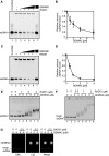
Figure 1.
Figure 1.
GEMIN2 stimulates RAD51–DNA filament formation. (A) Polyacrylamide gel electrophoresis to examine the formation of the RAD51–DNA filament. RAD51 (4 µM) and GEMIN2 were incubated with 10 µM 49-mer ssDNA. DNA was visualized by SYBR Gold (Invitrogen) staining. The GEMIN2 concentrations were 0 µM (lane 2), 1 µM (lane 3), 2 µM (lane 4), 4 µM (lane 5) and 8 µM (lanes 6 and 7). Under these experimental conditions, 90% of the input ssDNA was estimated as being in the RAD51-bound fraction in the absence of the GEMIN2 protein. (B) Quantification of experiments shown in panel A. The amounts of complexes formed were estimated from the residual free DNA substrates, and unbound ssDNA fractions relative to lane 2 of panel A were plotted. Average values of three independent experiments are shown with standard deviation values. (C) Polyacrylamide gel electrophoresis, as in panel A. RAD51 (2 µM) and GEMIN2 were incubated with 6 µM 49-mer dsDNA. DNA was visualized by SYBR Gold (Invitrogen) staining. The GEMIN2 concentrations were 0 µM (lane 2), 0.5 µM (lane 3), 1 µM (lane 4), 2 µM (lane 5) and 4 µM (lanes 6 and 7). (D) Quantification of experiments shown in panel C. The amounts of complexes formed were estimated from the residual free DNA substrates, and unbound dsDNA fractions relative to lane 2 of panel C were plotted. Average values of three independent experiments are shown with standard deviation values. (E) Agarose gel electrophoresis to examine the formation of the RAD51-ssDNA filament. RAD51 was incubated in the presence or absence of the GEMIN2 protein, followed by addition of ϕX174 ssDNA (20 µM). DNA was visualized by ethidium bromide staining. (F) Agarose gel electrophoresis to examine the formation of the RAD51–dsDNA filament. RAD51 was incubated in the presence or absence of the GEMIN2 protein, followed by addition of linear ϕX174 dsDNA (10 µM). Results presented as in panel E. (G) Agarose gel electrophoresis to assess the complex formation between the RAD51-dsDNA filament and GEMIN2. GEMIN2 was labeled with Cy5 and dsDNA was stained with EtBr. Note that GEMIN2 facilitated the formation of the RAD51-dsDNA filament, but did not bind to the filament.
Figure 2.
Figure 2.
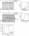
GEMIN2 stabilizes the RAD51–DNA filament. (A) Complex formation of RAD51 and dsDNA was evaluated by electrophoresis of unbound free DNA in agarose gel. Increased concentrations of competitor DNA were incubated with 2 µM of RAD51 in the presence or absence of 4 µM of GEMIN2, prior to the addition of ϕX174 dsDNA. (B) Quantification of results from panel A. The relative amounts of RAD51-unbound DNA are shown. Closed and open circles indicate experiments with and without GEMIN2. Average values and standard deviation were calculated from three independent experiments. (C) Complex formation of RAD51 and dsDNA in the presence of the BRC4 polypeptide. The experiments were done as described for panel A. (D) Quantification of the data from panel C. (E) Surface plasmon resonance analysis. The RAD51- or GEMIN2-conjugated sensor chips were used. Sensorgrams of RAD51-BRC4 and GEMIN2-BRC4 interactions are presented. The BRC4 polypeptide concentration was 10 µM. Time 0 of the horizontal axis indicates the initiation time of the peptide injection.
Figure 3.
Figure 3.
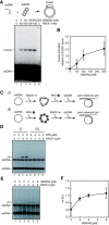
GEMIN2 enhances the homologous-pairing and strand-exchange activities of RAD51. (A) GEMIN2 stimulates the RAD51-mediated homologous pairing. RAD51 and GEMIN2 were incubated at 37°C for 5 min. After this incubation, a 32P-labeled 50-mer oligonucleotide (1 µM) was added, and the samples were further incubated at 37°C for 5 min. The reactions were then initiated by the addition of the pB5Sarray superhelical dsDNA (20 µM), and were continued at 37°C for 30 min. The reactions were stopped by the addition of SDS and proteinase K, and the deproteinized reaction products were separated by 1% agarose gel electrophoresis in 1× TAE buffer. The gels were dried, exposed to an imaging plate and visualized using an FLA-7000 imaging analyzer (Fujifilm, Tokyo, Japan). The reactions were conducted with 100 nM RAD51 in the presence of increasing amounts of GEMIN2. A schematic representation of the homologous pairing is presented on the top of the panel. (B) Graphic representation of the experiments shown in panel A. Amounts of D-loops relative to that of the RAD51 alone are plotted. The average values of three independent experiments are shown with the SD values. (C) Schematic representations of strand-exchange reactions. (i) The RAD51-ssDNA complexes are formed before the RPA addition. (ii) The RPA-ssDNA complexes are formed before the RAD51 addition. (D) Strand-exchange reactions where RPA was added to ϕX174 circular ssDNA (20 µM), after [lanes 1–4, panel C(i)] or before [lanes 5–8, panel C(ii)] incubation of the ssDNA with RAD51. Strand-exchange reactions were initiated by the addition of ϕX174 linear dsDNA (20 µM) and (NH4)2SO4 (100 mM), and incubated for 30 min. The deproteinized products of the reaction mixtures were separated using 1% agarose gel electrophoresis and were visualized by SYBR Gold (Invitrogen) staining. (E) GEMIN2 enhances strand exchange. ssDNA was incubated with RPA and then with RAD51 [panel C(ii)]. The indicated amounts of GEMIN2 were pre-incubated with RAD51, and subsequently added to the reaction mixture containing the ssDNA and RPA. (F) Quantification of panel E. The band intensities of the joint molecule (jm) products were quantified as the percentage of the entire input of the ssDNA and dsDNA molecules. Average values of three independent experiments are shown with standard deviation values.
Figure 4.
Figure 4.
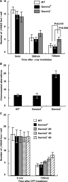
Defective repair of DSBs induced by γ-rays and camptothecin in GEMIN2-deficient cells. (A) Time line of γ-H2AX foci per cell at the indicated time after treatment with 4Gy γ-rays. Results for WT cells and for GEMIN2−/−tetGEMIN2 untreated (GEMIN2+) and treated (GEMIN2) with doxycycline for 4 days are shown. Error bars represent standard deviation. Statistical analysis was performed using the t test. (B) Ionizing-radiation-induced chromosome aberrations. Mitotic cells were harvested at 6 h after exposure of an asynchronous population of cells to 4Gy γ-rays. Irradiated cells were treated with colcemid for the last 3 h before harvest to enrich the mitotic cells. Results from WT and GEMIN2−/−tetGEMIN2 cells untreated with doxycycline (GEMIN2+) and treated (GEMIN2) with doxycycline for 4 days are shown. The number of chromosomal aberrations per 50 mitotic cells is shown on the Y-axis. Error bars represent standard deviation. (C) Time course of the formation of γ-H2AX foci per cell at the indicated times after treatment with camptothecin (CPT, 100 ng). Results for WT cells and for GEMIN2−/−tetGEMIN2 untreated (GEMIN2+) and treated (2 days, GEMIN2 2D; 3 days, GEMIN2 3D; 4 days, GEMIN2 4D) with doxycycline are shown. CPT is supposed to induce DSBs only in cells at the S phase, and the number of γ-H2AX foci was counted only in γ-H2AX foci positive cells. Error bars represent standard deviation.
Figure 5.
Figure 5.
HR-mediated DSB repair is impaired in GEMIN2-deficient cells. (A) Schematic of the HR-dependent DSB repair assay. Transient expression of the I-SceI restriction enzyme generates a specific DSB in an artificial HR substrate, DR-GFP, inserted into the endogenous OVALBUMIN locus. The SceGFP gene does not produce functional green fluorescent protein (GFP) because of a frame-shift mutation at the I-SceI recognition sequences. Functional GFP is produced only when the recognition sequences are eliminated by HR with the downstream internal GFP fragment (iGFP). (B) Distribution of GEMIN2−/−tetGEMIN2 cells with or without GFP expression. The population of the cells for GFP expression was measured at 12 h after transfection of I-SceI-expression plasmid [I-Sce(+)] or control plasmid [I-Sce(−)] in GEMIN2−/−tetGEMIN2 cells untreated (GEMIN2+) and treated (GEMIN2) with doxycycline for 3 days. (C) Percentage of cells expressing GFP in the indicated cell line was calculated. Data shown are the mean of three experiments. Error bars indicate standard deviation. (D) Transient transfection efficiency of an intact GFP expression plasmid in the indicated cell lines. Error bars indicate standard deviation.
Figure 6.
Figure 6.
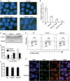
IR-induced Rad51 subnuclear foci are decreased in the absence of GEMIN2. (A) Immunostaining of irradiated WT and GEMIN2−/−tetGEMIN2 cells with the Rad51 antibody. Cells were fixed 3 h after irradiation of 4Gy γ-ray. GEMIN2−/−tetGEMIN2 cells untreated (GEMIN2+) and treated (GEMIN2, 2 or 4 days) with doxycycline are shown. (B) Quantification of Rad51 foci in individual cells of the indicated genotype. Data shown are the means of three experiments. Error bars indicate standard deviation. (C) U2OS cells were transfected with siRNA oligos specific to GEMIN2 or with control siRNA. Cell extract was prepared at 48 h and immunoblotted as indicated. (D) Cell-cycle distribution 48 h after addition of control (GFP) and GEMIN2-specific siRNAs. Cells were pulse-labeled with BrdU for 10 min and subsequently stained with FITC-conjugated anti-BrdU antibody (Y-axis, log scale) and propidium iodide (PI) (X-axis, linear scale). The upper gate indicates cells incorporating BrdU (S-phase); the lower middle gate indicates G1 cells; and the lower right gate displays G2/M cells. The sub G1 fraction (lower left gates) indicates dead cells. The numbers given in each gate indicate the percentages of gated events. (E) Rad51 focus formation in human cells depleted of GEMIN2 by siRNA. Cells were immunostained 3 h after 4Gy IR exposure. The histogram shows the percentage of cells displaying less than 6 RAD51 foci, 6 to 10 foci, and more than 10 foci per cell. >100 cells were analyzed for each data point. (F) FK2-conjugated ubiquitin focus formation following IR exposure is normal in GEMIN2-depleted DT40 cells. GEMIN2 depletion was performed by treating GEMIN2−/−tetGEMIN2 cells with doxycycline for 4 days. Error bars indicate standard deviation. (G) The RPA ssDNA binding protein normally accumulates at DSBs 60 min after exposure to a 405 nm pulse laser in HeLa cells treated with siRNA against GEMIN2 or control siRNA for 48 h.

Similar articles

Cited by

References

    1. Pommier Y. Topoisomerase I inhibitors: camptothecins and beyond. Nat. Rev. Cancer. 2006;6:789–802. - PubMed
    1. Sung PA, Libura J, Richardson C. Etoposide and illegitimate DNA double-strand break repair in the generation of MLL translocations: new insights and new questions. DNA Repair. 2006;5:1109–1118. - PubMed
    1. Sonoda E, Hochegger H, Saberi A, Taniguchi Y, Takeda S. Differential usage of non-homologous end-joining and homologous recombination in double strand break repair. DNA Repair. 2006;5:1021–1029. - PubMed
    1. Takeda S, Nakamura K, Taniguchi Y, Paull TT. Ctp1/CtIP and the MRN complex collaborate in the initial steps of homologous recombination. Mol. Cell. 2007;28:351–352. - PubMed
    1. Sonoda E, Sasaki MS, Buerstedde JM, Bezzubova O, Shinohara A, Ogawa H, Takata M, Yamaguchi-Iwai Y, Takeda S. Rad51-deficient vertebrate cells accumulate chromosomal breaks prior to cell death. EMBO J. 1998;17:598–608. - PMC - PubMed

Publication types