Review
. 2010 May 27;365(1546):1481-500.
doi: 10.1098/rstb.2009.0294.

Testicular postgenomics: targeting the regulation of spermatogenesis

Affiliations
Review

Testicular postgenomics: targeting the regulation of spermatogenesis

Pierre Calvel et al. Philos Trans R Soc Lond B Biol Sci. .

Abstract

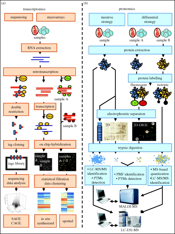
Sperm are, arguably, the most differentiated cells produced within the body of any given species. This is owing to the fact that spermatogenesis is an intricate and highly specialized process evolved to suit the individual particularities of each sexual species. Despite a vast diversity in method, the aim of spermatogenesis is always the same, the idealized transmission of genetic patrimony. Towards this goal certain requirements must always be met, such as a relative twofold reduction in ploidy, repackaging of the chromatin for transport and specialized enhancements for cell motility, recognition and fusion. In the past 20 years, the study of molecular networks coordinating male germ cell development, particularly in mammals, has become more and more facilitated thanks to large-scale analyses of genome expression. Such postgenomic endeavors have generated landscapes of data for both fundamental and clinical reproductive biology. Continuous, large-scale integration analyses of these datasets are undertaken which provide access to very precise information on a myriad of biomolecules. This review presents commonly used transcriptomic and proteomic workflows applied to various testicular germ cell studies. We will also provide a general overview of the technical possibilities available to reproductive genomic biologists, noting the advantages and drawbacks of each technique.


Figures

Figure 1.
Figure 1.
The basics of the transcriptomic and proteomic technical workflows. (a) Transcriptomics: the high-scale analysis of gene expression can be operated through two distinct strategies. In sequencing-based approaches such as SAGE and CAGE, small DNA fragments (10–14 bp) are generated from cDNAs and subcloned in a DNA library, in which the abundance of each tag is correlated to the level of expression of the corresponding gene. The high-throughput sequencing of libraries corresponding to various samples gives access to quantitative data on genes expression. On the other hand, oligonucleotide microarrays are based on the ability of complementary DNA or RNA fragments to hybridize together. Two classes of arrays are frequently used: in spotted DNA microarrays, micro droplets of long complementary DNA fragments are spotted on a chip. The cDNAs prepared from two samples are labelled with fluorescent dyes and hybridized simultaneously on the chip. The ratio of the two fluorescences emitted by each spot will determine the relative expression of the corresponding gene in the two samples. With the in situ synthesized microarrays, two sets of 11 25 bp-probes are directly synthesized for each gene on the chip. For each sample, biotinylated cRNAs are produced from cDNAs and hybridized individually on the chip. The RNAs' abundance is revealed by a peroxydase assay. (b) Proteomics: the first strategy dealing with the high-throughput analysis of proteins aims to systematically identifying the protein content of a biological sample. Classically, proteins extracted from a whole organ or isolated cells are separated by two-dimensional-gel electrophoresis. Each detectable spot is then excised from the gel and submitted to a trypsic digestion. The resulting peptides are listed by MALDI-MS mass spectrometry and the corresponding protein is identified thanks to the peptide mass fingerprint (PMF) technique. Recently, the progress made with highly sensitive mass spectrometry enabled scientists to avoid the complex two-dimensional separation process. A whole trypsic-digested proteic sample can thus be separated by liquid chromatography and directly injected in an ESI-MS, where the peptides will be identified by tandem mass spectrometry (MS/MS). An intermediate way is to prefractionnate the samples through one-dimensional-gel electrophoresis. Bands containing hundreds of proteins are then sliced off the gel and trypsin-treated before their identification by LC-ESI-MS/MS sequencing. Quantification can also be considered with proteomics, either by electrophoresis-based or MS-based methods. Following technical principles similar to those used in spotted oligonucleotide microarrays, the two-dimensional difference in gel electrophoresis (2D-DIGE) relies on the simultaneous analysis of two differentially cyanine-labelled samples. Protein spot exhibiting asymmetrical fluorescence can be identified by PMF. Differential analysis can also be performed with quantitative mass spectrometry. In the case of isotope-coded protein label technique (ICPL), samples can be differentially labelled with light or heavy isotopes and digested together prior to MS analysis. The determination of the ratio between the intensity of a light and heavy labelled peak on the mass spectra followed by MS/MS protein identification allows the determination of the protein relative expression level within the two samples. Other quantification techniques based on MS analysis are available (ICAT, ITRAQ), but they require post-trypsic digestion labelling procedures.
Figure 2.
Figure 2.
Pathways for the self-renewal and differentiation of spermatogonial stem cells (SSC). The daily production of millions of spermatozoa in mammals is ensured by the presence within the male germ line of stem cells able to maintain their own stock and to differentiate and continuously initiate new waves of spermatogenesis. The balance between the maintenance of spermatogonial stem cells, their proliferation and their differentiation into mature spermatogonia is triggered by intrinsic factors, such as PLZF, and by signals from the spermatogonial stem cell niche, including Sertolian signals, such as GDNF, ERM or Kit ligand. GDNF: glial cell line-derived neurotrophic factor; ERM: Ets-related molecule; PLZF: promyelocytic leukemia zinc finger. References: 1, Chen et al. (2005); 2, Hofmann et al. (2005); 3, Oatley et al. (2006); 4, Kokkinaki et al. (2009); 5, Schmidt et al. (2009); 6, Hamra et al. (2004); 7, Costoya et al. (2004); 8, Guillaume et al. (2000); 9, Com et al. (2003); 10, Rossi et al. (2008); 11, Zhou et al. (2008).
Figure 3.
Figure 3.
Deciphering spermatogenesis through expression analyses. Spermatogenesis is a complex, coordinated and continuous process by which spermatogonia give rise to mature spermatozoa. In rodents, the first wave of this process starts a few days after birth, when gonocytes (prespermatogonia) resume proliferation and become undifferentiated spermatogonia or spermatogonial stem cells. Once SSC are engaged in differentiation process (i.e. they become differentiated spermatogonia) they undergo six successive mitotic divisions, giving rise to meiotic spermatocytes, which go through two consecutive divisions, with a single round of DNA replication, to give rise to haploid spermatids. The final step in spermatogenesis consists of the transformation of spermatids into spermatozoa and is called spermiogenesis. References: 1, Divina et al. (2005); 2, Shima et al. (2004); 3, Almstrup et al. (2004); 4, Ellis et al. (2004); 5, Clemente et al. (2006); 6, Schultz et al. (2003); 7, Yao et al. (2004); 8, Fox et al. (2003); 9, Huang et al. (2005); 10, Zhu et al. (2006); 11, Cagney et al. (2005); 12, Ro et al. (2007); 13, Yan et al. (2009); 14, Yan et al. (2007); 15, Tamminga et al.(2008); 16, Govin et al. (2006); 17, Wu et al. (2004); 18, Namekawa et al. (2006); 19, Chalmel et al. (2007b); 20, Schlecht et al. (2004); 21, Rolland et al. (2007).
Figure 4.
Figure 4.
The main components and maturation of spermatozoa. Once released from the seminiferous epithelium, spermatozoa are transferred to the epididymis. During their transit in the epididymis, spermatozoa become motile. However, they are not yet competent for fertilization. Within the female reproductive tract, they finally acquire the ability to fertilize eggs, during a time-dependent process called capacitation, making it possible for them to undergo the acrosomal reaction and to bind to the ovocyte membrane. References: 1, Ostermeier et al. (2002); 2, Ostermeier et al. (2005); 3, Wang et al. (2004); 4, Nguyen et al. (2008); 5, Zhao et al. (2006); 6, Dorus et al. (2006); 7, Baker et al. (2008b); 8, Cao et al. (2006); 9, Krisfalusi et al. (2006); 10, Stein et al. (2006); 11, Baker et al. (2008a); 12, Peddinti et al. (2008); 13, Martinez-Heredia et al. (2006); 14, Johnston et al. (2005); 15, Baker et al. (2007); 16, Kim et al. (2006); 17, Baker et al. (2005); 18, Sleight et al. (2005); 19, Nixon et al. (2009); 20, Lefievre et al. (2007); 21, Ficarro et al. (2003); 22, Lalancette et al. (2006); 23, Platt et al. (2009); 24, Zhao et al. (2007); 25, Martinez-Heredia et al. (2008); 26, de Mateo et al. (2007); 27, Bohring et al. (2001); 28, Bohring & Krause (2003); 29, Fijak et al. (2005); 30, Paradowska et al. (2006); 31, Domagala et al. (2007).
Figure 5.
Figure 5.
Data mining: going beyond expression profiles. Figure adapted from Stein et al. (2006). (a) Filtering strategy. Cross-species comparison and tissue profiling identified 80 testis-specific orthologues with conserved expression patterns in mouse, rat and human spermatogenesis. Genes differentially expressed during spermatogenesis were identified for each species. Differentially expressed genes corresponding to orthologues represented on microarrays for all three species were identified, using the HomoloGene database. Finally, expression data for 17 mouse somatic tissues downloaded from the GEO web server were used to select testis-specific genes. SE, SPG, SC, ST, TU and TT correspond to Sertoli cell, spermatogonia, pachytene spermatocyte, early spermatid, seminiferous tubule and total testis samples, respectively. SO, MI, MEI and PM correspond to the somatic, mitotic, meiotic and postmeiotic expression clusters, respectively. (b) Promoter analysis. Automated multi-step promoter analysis showed mouse testis-specific postmeiotic gene promoter regions to be specifically enriched in the CRE motif. We show here the results for Capza3, a gene also identified as present within the interaction node of conserved and testis-specific genes. (c) Interaction network analysis. Data from IntAct, MINT and BioGRID were used to monitor interactions between conserved and testis-specific gene products. Some proteins important for male reproduction were found within these networks and were found to associate with a large number of interacting factors. As not all the interacting factors were detected on microarray analyses, interaction network analysis might help to extend expression data.

Similar articles

Cited by

References

    1. Aebersold R., Mann M.2003Mass spectrometry-based proteomics. Nature 422, 198–207 (doi:10.1038/nature01511) - DOI - PubMed
    1. Almstrup K., Nielsen J. E., Hansen M. A., Tanaka M., Skakkebaek N. E., Leffers H.2004Analysis of cell-type-specific gene expression during mouse spermatogenesis. Biol. Reprod. 70, 1751–1761 (doi:10.1095/biolreprod.103.026575) - DOI - PubMed
    1. Anderson N. L., Anderson N. G.1998Proteome and proteomics: new technologies, new concepts, and new words. Electrophoresis 19, 1853–1861 (doi:10.1002/elps.1150191103) - DOI - PubMed
    1. Andrabi S. M.2007Mammalian sperm chromatin structure and assessment of DNA fragmentation. J. Assist. Reprod. Genet. 24, 561–569 (doi:10.1007/s10815-007-9177-y) - DOI - PMC - PubMed
    1. Ashburner M., et al. 2000Gene ontology: tool for the unification of biology. The gene ontology consortium. Nat. Genet. 25, 25–29 - PMC - PubMed

LinkOut - more resources