Review
. 2010 May 27;365(1546):1557-69.
doi: 10.1098/rstb.2009.0258.

Non-classical actions of testosterone and spermatogenesis

Affiliations
Review

Non-classical actions of testosterone and spermatogenesis

William H Walker. Philos Trans R Soc Lond B Biol Sci. .

Abstract

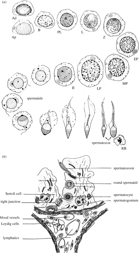
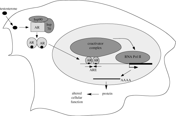
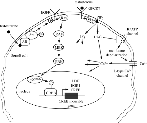
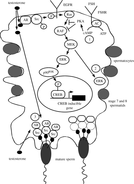
Testosterone is essential to maintain spermatogenesis and male fertility. In the absence of testosterone stimulation, spermatogenesis does not proceed beyond the meiosis stage. After withdrawal of testosterone, germ cells that have progressed beyond meiosis detach from supporting Sertoli cells and die, whereas mature sperm cannot be released from Sertoli cells resulting in infertility. The classical mechanism of testosterone action in which testosterone activates gene transcription by causing the androgen receptor to translocate to and bind specific DNA regulatory elements does not appear to fully explain testosterone regulation of spermatogenesis. This review discusses two non-classical testosterone signalling pathways in Sertoli cells and their potential effects on spermatogenesis. Specifically, testosterone-mediated activation of phospholipase C and calcium influx into Sertoli cells is described. Also, testosterone activation of Src, EGF receptor and ERK kinases as well as the activation of the CREB transcription factor and CREB-mediated transcription is reviewed. Regulation of germ cell adhesion to Sertoli cells and release of mature sperm from Sertoli cells by kinases regulated by the non-classical testosterone pathway is discussed. The evidence accumulated suggests that classical and non-classical testosterone signalling contribute to the maintenance of spermatogenesis and male fertility.


Figures

Figure 1.
Figure 1.
(a) The process of spermatogenesis. A type spermatogonia undergo mitotic divisions to become B spermatogonia which then divide and differentiate into prelepotene spermatocytes (PL) that then become leptotene (L), zygotene (Z) and then early mid and late pachytene spematocytes (EP, MP, LP). After the first meiotic division, secondary spermatocytes are formed (II) that then divide again resulting in haploid round spematids. The spermatids elongate, shed much of their cytoplasm as residual bodies (RB) and differentiate until mature spermatozoa are formed. (b) The anatomy of the testis. The outline of three seminiferous tubules is shown surrounding the interstitial area containing blood vessels, lymphatic tissue and colonies of Leydig cells. The seminiferous tubules are surrounded by basement membrane and myoid peritubullalr cells. Spermatogonia are located on the basement membrane. Spermatocytes and spermatids and mature spermatozoa are located apical to the tight junctions between Sertoli cells that make up the blood testis barrier. Sertoli cells extend from the basement membrane to the lumen of the tubule.
Figure 2.
Figure 2.
The classical testosterone signalling pathway. Testosterone diffuses through the plasma membrane and binds with the AR. The AR undergoes an alteration in conformation allowing it to be released from heat shock proteins in the cytoplasm. AR then is able to translocate to the nucleus where it binds to specific DNA sequences called androgen response elements (AREs). AR binding to an ARE allows the recruitment of co-activator and co-repressor proteins that alter the expression of genes to alter cellular function.
Figure 3.
Figure 3.
Non-classical testosterone signalling pathways. Left side: testosterone interacts with the classical AR which then is able to recruit and activate Src, which causes the activation of the EGF receptor via an intracellular pathway. The EGF receptor then activates the MAP kinase cascade most probably through Ras, resulting in the sequential activation of RAF and MEK and then ERK that activates p90-kinase, which is known to phosphorylate CREB on serine 133. As a result, CREB-regulated genes such as lactate dehydrogenase A (LDH-A) and early growth response 1 (EGR1) and CREB can be induced by testosterone. Right side: Testosterone interacts with a receptor in the plasma membrane that has characteristics of a Gq coupled G-protein coupled receptor (GPCR). Phospholipase C (PLC) is activated to cleave PIP2 into IP3 and DAG. Lower concentrations of PIP2 inhibit K+ATP channels causing membrane depolarization and Ca2+ entry via L-type Ca2+ channels.
Figure 4.
Figure 4.
Regulation of spermatogenesis processes by non-classical testosterone signalling. The non-classical signalling pathway resulting in Src, ERK and CREB activation is shown for a stage VII–VIII seminiferous tubule cross section. (1) FSH-mediated increases in cAMP and PKA activity result in blocking of the non-classical testosterone signalling pathway at the Raf kinase step so that p-ERK levels would be restricted except for stages VII–VIII when FSHR levels are lowest and AR expression is highest in the Sertoli cell. (2) ERK activation contributes to testosterone-mediated Sertoli–germ cell adhesion and ERK activation may be particularly important during stages VII–VIII to remodel the connections between Sertoli cells and spermatids as they begin to elongate. (3) Testosterone entering the Sertoli cell from the interstitial space or the tubule lumen acts through Src kinase to promote the release of mature sperm from the Sertoli ES, most probably by Src phosphorylation of adaptor proteins (filled round circles) that anchor the cell–cell adhesion proteins (represented by lines extending from Sertoli cell to mature sperm) to actin filaments.

Similar articles

Cited by

References

    1. Awoniyi C. A., Santulli R., Sprando R. L., Ewing L. L., Zirkin B. R.1989Restoration of advanced spermatogenic cells in the experimentally regressed rat testis: quantitative relationship to testosterone concentration within the testis. Endocrinology 124, 1217–1223 (doi:10.1210/endo-124-3-1217) - DOI - PubMed
    1. Beardsley A., O'Donnell L.2003Characterization of normal spermiation and spermiation failure induced by hormone suppression in adult rats. Biol. Reprod. 68, 1299–1307 (doi:10.1095/biolreprod.102.009811) - DOI - PubMed
    1. Benten W. P. M., Benten M., Lieberherr M., Stamm O., Wrehlke C., Guo Z., Wunderlich F.1999Testosterone signaling through internalizable surface receptors in androgen receptor-free macrophages. Mol. Biol. Cell 10, 3113–3123 - PMC - PubMed
    1. Bremner W. J., Millar M. R., Sharpe R. M., Saunders P. T. K.1994Immunohistochemical localization of androgen receptors in the rat testis: evidence for stage-dependent expression and regulation by androgens. Endocrinology 135, 1227–1234 (doi:10.1210/en.135.3.1227) - DOI - PubMed
    1. Chang C., Chen Y. T., Yeh S. D., Xu Q., Wang R. S., Guillou F., Lardy H., Yeh S.2004Infertility with defective spermatogenesis and hypotestosteronemia in male mice lacking the androgen receptor in Sertoli cells. Proc. Natl Acad. Sci. USA 101, 6876–6881 (doi:10.1073/pnas.0307306101) - DOI - PMC - PubMed

Publication types

LinkOut - more resources