

Review
doi: 10.1098/rstb.2010.0026.
Spermatogonial stem cell regulation and spermatogenesis
Affiliations
- PMID: 20403877
- PMCID: PMC2871929
- DOI: 10.1098/rstb.2010.0026
Item in Clipboard
Review
Spermatogonial stem cell regulation and spermatogenesis
Philos Trans R Soc Lond B Biol Sci.
.
Abstract
This article will provide an updated review of spermatogonial stem cells and their role in maintaining the spermatogenic lineage. Experimental tools used to study spermatogonial stem cells (SSCs) will be described, along with research using these tools to enhance our understanding of stem cell biology and spermatogenesis. Increased knowledge about the biology of SSCs improves our capacity to manipulate these cells for practical application. The chapter concludes with a discussion of future directions for fundamental investigation and practical applications of SSCs.
Figures
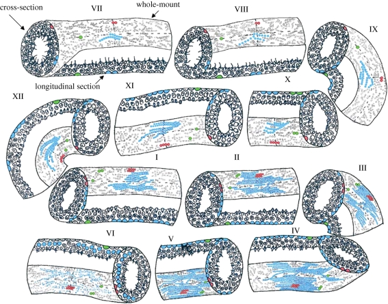
Mouse spermatogenic clone development by stage. The mouse spermatogenic cycle contains twelve stages (I–XII). Each stage is temporally unique, and the stages in the diagram represent the relative time each stage lasts in the mouse. Each stage in the diagram is shown in cross-sectional, longitudinal and whole-mount perspectives (labelled in stage VII). Three putative spermatogonial clones are highlighted in blue, red and green. The dotted lines in the whole-mount perspective indicate the planes of the cross section and longitudinal section views. For example, in stage VII, the red cell is in the vertical line and therefore appears in the cross-sectional view. A green cell is in the horizontal line, so is observed in the longitudinal section view. The development of three putative clones (blue, red and green) through one cycle of the seminiferous epithelium is shown. Stage VII: Aal-16 (blue); Apair (red); Asingle (green); stage VIII: A1 (clone of 16) (blue); Apair (red); Asingle (green); stage IX: A2 (clone of 32) (blue); Apair (red); Asingle (green); stage X: A2 (clone of 32) (blue); Aal-4 (red); Apair (green); stage XI: A3 (clone of 64) (blue); Aal-4 (red); Asingle (x2) (green); stage XII: A3 (clone of 64) (blue); Aal-4 (red); Asingle (x2) (green); stage I: A4 (clone of 128) (blue); Aal-8 (red); Asingle and Apair (green); stage II: intermediate spermatogonia (clone of 256) (blue); Aal-8 (red); Asingle and Apair (green); stage III: intermediate spermatogonia (clone of 256) (blue); Aal-8 (red); Asingle and Apair (green); stage IV: Type B Spermatagonia (clone of 512) (blue); Aal-8 (red); Asingle and Apair (green); stage V: Type B Spermatagonia (clone of 512) (blue); Aal-8 (red); Asingle and Apair (green); stage VI: primary spermatocytes (lifting off the basement membrane) (blue); Aal-8 (red); Asingle and Apair (green).
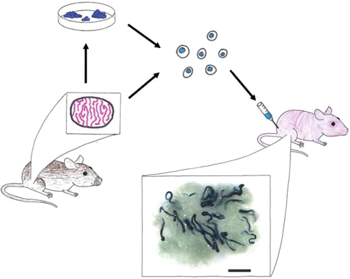
Spermatogonial stem cell (SSC) transplant assay. The functional analysis of SSCs is a retrospective assay of spermatogenic function. In this example, cells are isolated from a lacZ donor mouse testis and digested to produce a single cell suspension. Cells can then be maintained in culture or injected into the testes of an infertile recipient mouse. Recipient testes are typically analysed two to three months after transplantation for donor spermatogenesis (blue colonies in this example). A typical recipient testis is shown with blue colonies of donor-derived spermatogenesis (scale bar, 2 mm).

Genes expressed by stem, progenitor and differentiating spermatogonia. The As, seen at the top of the diagram, is responsible for self renewal and differentiation. Self-renewal is represented here by the Apair dividing to form two As. Differentiation is indicated by colour change (from dark to light) and the lengthening chain of germ cells. Genes are listed with their expression at the given stages of spermatogonial development. While stem cell activity is considered to reside in the pool of As spermatogonia, the tapered triangle on the left indicates that stem cell activity may extend to Apr and some Aal spermatogonia.
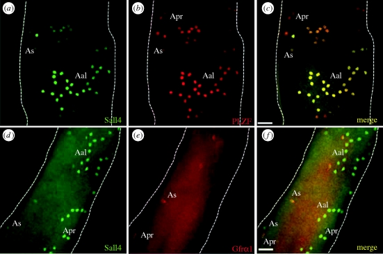
Immunofluorescent co-staining of adult mouse whole-mount seminiferous tubules. (a) SALL4 labels undifferentiated As, Apr and Aal spermatogonia. (b) PLZF labels undifferentiated As, Apr and Aal spermatogonia. (c) Merged picture from (a,b). SALL4 and PLZF are mostly co-expressed in undifferentiated spermatogonia. (d–f) Co-staining of SALL4 and GFRα1 reveals heterogeneity within the population of undifferentiated spermatogonia. GFRα1 expression appears more restricted than SALL4 or PLZF. Scale bar, 50 µm.
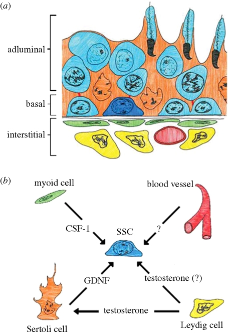
SSC niche. The SSC (dark blue) is diagrammed in its physical niche (a) surrounded by Sertoli cells (orange) and differentiating germ cells (light blue) within the seminiferous tubule. Niche components outside the tubule itself include myoid cells (green), blood vessels (red) and Leydig cells (yellow). The components of the niche and the some factors known to be provided by each are shown in (b). While some factors are known to act directly on the SSC, such as GDNF, others, like testosterone are important for spermatogenesis but may not act on the SSC.
References
-
- Anderson R., Schaible K., Heasman J., Wylie C.1999Expression of the homophilic adhesion molecule, Ep-CAM, in the mammalian germ line. J. Reprod. Fertil. 116, 379–384 - PubMed
-
- Antonangeli F., Giampietri C., Petrungaro S., Filippini A., Ziparo E.2009Expression profile of a 400-bp Stra8 promoter region during spermatogenesis. Microsc. Res. Tech. 72, 816–822 - PubMed
-
- Arregui L., Rathi R., Megee S. O., Honaramooz A., Gomendio M., Roldan E. R., Dobrinski I.2008Xenografting of sheep testis tissue and isolated cells as a model for preservation of genetic material from endangered ungulates. Reproduction 136, 85–93 (doi:10.1530/REP-07-0433) - DOI - PubMed
-
- Ballow D., Meistrich M. L., Matzuk M., Rajkovic A.2006aSohlh1 is essential for spermatogonial differentiation. Dev. Biol. 294, 161–167 (doi:10.1016/j.ydbio.2006.02.027) - DOI - PubMed
-
- Ballow D. J., Xin Y., Choi Y., Pangas S. A., Rajkovic A.2006bSohlh2 is a germ cell-specific bHLH transcription factor. Gene Expr. Patterns 6, 1014–1018 (doi:10.1016/j.modgep.2006.04.007) - DOI - PubMed
Publication types
MeSH terms
Grants and funding
LinkOut - more resources
Full Text Sources
Other Literature Sources
Medical