. 2010 May;133(Pt 5):1415-27.
doi: 10.1093/brain/awq078. Epub 2010 Apr 19.

Clinical and imaging heterogeneity of polymicrogyria: a study of 328 patients

Affiliations

Clinical and imaging heterogeneity of polymicrogyria: a study of 328 patients

Richard J Leventer et al. Brain. 2010 May.

Abstract

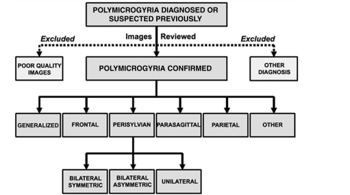
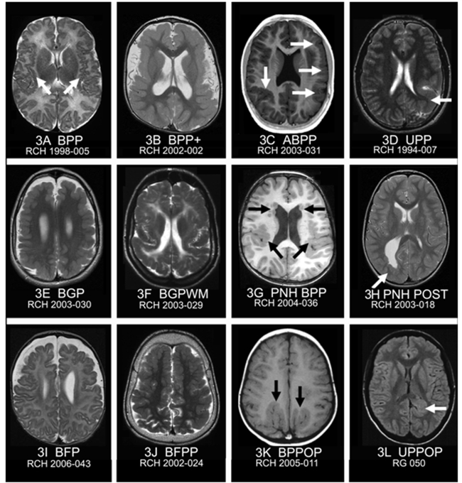
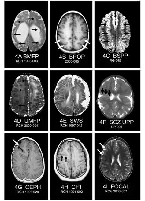
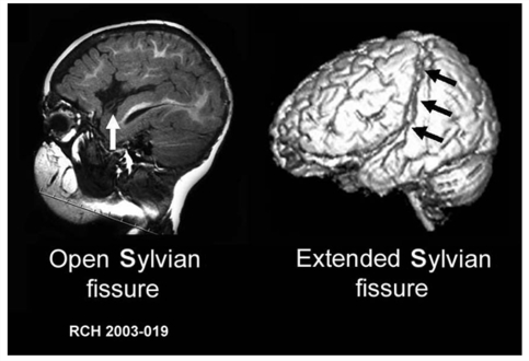
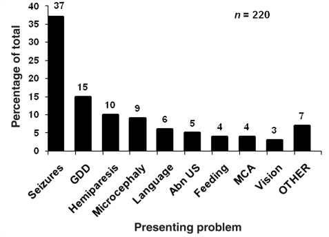
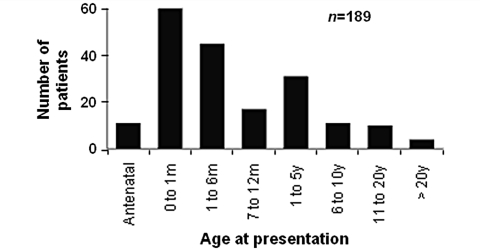
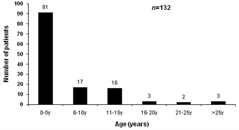
Polymicrogyria is one of the most common malformations of cortical development and is associated with a variety of clinical sequelae including epilepsy, intellectual disability, motor dysfunction and speech disturbance. It has heterogeneous clinical manifestations and imaging patterns, yet large cohort data defining the clinical and imaging spectrum and the relative frequencies of each subtype are lacking. The aims of this study were to determine the types and relative frequencies of different polymicrogyria patterns, define the spectrum of their clinical and imaging features and assess for clinical/imaging correlations. We studied the imaging features of 328 patients referred from six centres, with detailed clinical data available for 183 patients. The ascertainment base was wide, including referral from paediatricians, geneticists and neurologists. The main patterns of polymicrogyria were perisylvian (61%), generalized (13%), frontal (5%) and parasagittal parieto-occipital (3%), and in 11% there was associated periventricular grey matter heterotopia. Each of the above patterns was further divided into subtypes based on distinguishing imaging characteristics. The remaining 7% were comprised of a number of rare patterns, many not described previously. The most common clinical sequelae were epileptic seizures (78%), global developmental delay (70%), spasticity (51%) and microcephaly (50%). Many patients presented with neurological or developmental abnormalities prior to the onset of epilepsy. Patients with more extensive patterns of polymicrogyria presented at an earlier age and with more severe sequelae than those with restricted or unilateral forms. The median age at presentation for the entire cohort was 4 months with 38% presenting in either the antenatal or neonatal periods. There were no significant differences between the prevalence of epilepsy for each polymicrogyria pattern, however patients with generalized and bilateral forms had a lower age at seizure onset. There was significant skewing towards males with a ratio of 3:2. This study expands our understanding of the spectrum of clinical and imaging features of polymicrogyria. Progression from describing imaging patterns to defining anatomoclinical syndromes will improve the accuracy of prognostic counselling and will aid identification of the aetiologies of polymicrogyria, including genetic causes.


Figures

Figure 1
Figure 1
Key MRI features of polymicrogyria. The centre image shows perisylvian polymicrogyria. (A) Undulation and irregularity of the cortical surface along the Sylvian fissure. (B, C) Comparison of the thickness of normal cortex (B, 4 mm) with the apparent thickening of polymicrogyric cortex (C, 10 mm). (D) Stippling and irregularity at the grey–white junction.
Figure 2
Figure 2
Process for selection of polymicrogyria cases and initial classification.
Figure 3
Figure 3
MRI features of common patterns and subtypes of polymicrogyria. All images are T1, T2 or fluid attenuated inversion recovery axial. (A, B) Bilateral perisylvian polymicrogyria with polymicrogyria either limited to the perisylvian cortex (BPP), or extending beyond it (BPP+). (C) Asymmetric bilateral perisylvian polymicrogyria (ABPP) with polymicrogyria involving the posterior third of the right Sylvian fissure and the left Sylvian fissure along its entire length (arrows). (D) Unilateral perisylvian polymicrogyria (UPP) with polymicrogyria lining the left Sylvian fissure that is abnormally extended postero-superiorly. (E) Bilateral generalized polymicrogyria (BGP) showing no clear gradient or region of maximal severity. (F) Bilateral generalized polymicrogyria with abnormal white matter (BGPWM). Note abnormal high signal in the subcortical and deep white matter. (G) Bilateral periventricular grey matter heterotopia with bilateral perisylvian polymicrogyria (PNH BPP) (arrows). (H) Right-sided posterior periventricular grey matter heterotopia (arrow) associated with overlying polymicrogyria (PNH POST). (I) Bilateral frontal (only) polymicrogyria (BFP) with bilateral symmetric polymicrogyria involving the majority of the frontal lobes with abrupt cut-off in the mid-frontal regions. (J) Bilateral frontoparietal polymicrogyria (BFPP) with bilateral symmetric polymicrogyria involving the frontal lobes and extension posteriorly into the parietal lobes. (K) Bilateral parasagittal parieto-occipital polymicrogyria (BPPOP) with bilateral symmetric polymicrogyria lining abnormal gyri radiating antero-laterally from the parasagittal parieto-occipital region (arrows). (L) A mild form of unilateral parasagittal parieto-occipital polymicrogyria (UPPOP) with polymicrogyria lining a deep and abnormally-oriented sulcus in the left parasagittal region.
Figure 4
Figure 4
MRI features of atypical and rare patterns of polymicrogyria. All images are T1, T2 or fluid attenuated inversion recovery axial. (A) Bilateral multifocal polymicrogyria (BMFP) patchy throughout both hemispheres (arrows) with associated abnormal white matter signal and ventriculomegaly. (B) Bilateral parieto-occipital polymicrogyria (BPOP) with bilateral symmetric polymicrogyria in lateral parieto-occipital regions (arrows). (C) Bilateral superior parasagittal polymicrogyria (BSPP) with polymicrogyria lining abnormal deep symmetric parasagittal gyri extending from the frontal poles to the parietal lobes (arrows). (D) Unilateral multifocal polymicrogyria (UMFP) with multifocal polymicrogyria throughout the right hemisphere (arrows). This case was confirmed as polymicrogyria by pathology. (E) MRI from a child with Sturge–Weber syndrome who had epilepsy, a left-sided facial haemangioma and left eye glaucoma. The images show atrophy of the left hemisphere with extensive polymicrogyria. CT scanning showed cortical calcifications and contrast-enhanced imaging suggested pial angiomatosis typical of Sturge–Weber syndrome. (F) Unilateral closed-lip schizencephaly and contralateral perisylvian polymicrogyria (schizencephaly UPP). There is extensive polymicrogyria bilaterally, maximal along the Sylvian fissures. On both sides, there were deep clefts extending towards the lateral ventricles lined by polymicrogyria. On the right side, this cleft communicated with the lateral ventricle showing the ‘pia-ependymal seam’ typical of schizencephaly (arrows). The left-sided cleft did not communicate with the lateral ventricle. (G) Polymicrogyria associated with an encephalocoele (CEPH) showing a deep cleft (arrow) surrounded by irregular, thickened grey matter consistent with polymicrogyria underlying the site of a previously-repaired right frontal encephalocoele. (H) Polymicrogyria associated with a deep cleft (CFT) in the right mid-frontal lobe extending towards (but not communicating with) the lateral ventricle (arrows). This cleft is lined by irregular, thickened grey matter suggestive of polymicrogyria. (I) Focal polymicrogyria (FOCAL) with an irregular area of polymicrogyria over the right mid-frontal region (arrows).
Figure 5
Figure 5
MRI of ‘open’ and ‘extended’ Sylvian fissure patterns. Parasagittal T1 MRI (left) showing an ‘open’ Sylvian fissure with failure of apposition of the inferior frontal and the superior temporal gyri anteriorly (white arrow). 3D surface reconstruction of a patient with perisylvian polymicrogyria (right) showing an ‘extended’ Sylvian fissure, posterosuperiorly into the parietal lobe (black arrows).
Figure 6
Figure 6
Types and frequency of presenting problem expressed as a percentage of all presenting problems. GDD = global developmental delay; language = isolated language delay; Abn US = abnormal antenatal ultrasound; feeding = feeding difficulties; MCA = multiple congenital anomalies; vision = visual impairment.
Figure 7
Figure 7
Distribution of ages at presentation in 189 patients with polymicrogyria.
Figure 8
Figure 8
Age at seizure onset in 132 patients with polymicrogyria and epilepsy.

References

    1. Aicardi J. Aicardi syndrome. Brain Dev. 2005;27:164–71. - PubMed
    1. Barkovich AJ, Hevner R, Guerrini R. Syndromes of bilateral symmetrical polymicrogyria. Am J Neuroradiol. 1999;20:1814–21. - PMC - PubMed
    1. Barkovich AJ, Kuzniecky RI, Jackson GD, Guerrini R, Dobyns WB. A developmental and genetic classification for malformations of cortical development. Neurology. 2005;65:1873–87. - PubMed
    1. Chang BS, Piao X, Bodell A, Basel-Vanagaite L, Straussberg R, Dobyns WB, et al. Bilateral frontoparietal polymicrogyria: clinical and radiological features in 10 families with linkage to chromosome 16. Ann Neurol. 2003;53:596–606. - PubMed
    1. Chang BS, Piao X, Giannini C, Cascino GD, Scheffer I, Woods CG, et al. Bilateral generalized polymicrogyria (BGP): a distinct syndrome of cortical malformation. Neurology. 2004;62:1722–8. - PubMed

Publication types

MeSH terms