

Inhibitory neurones of the spinal substantia gelatinosa mediate interaction of signals from primary afferents
- PMID: 20403977
- PMCID: PMC2911212
- DOI: 10.1113/jphysiol.2010.188052
Inhibitory neurones of the spinal substantia gelatinosa mediate interaction of signals from primary afferents
Abstract
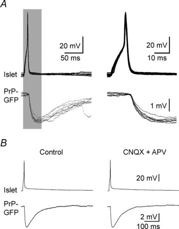
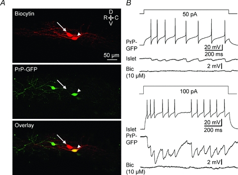
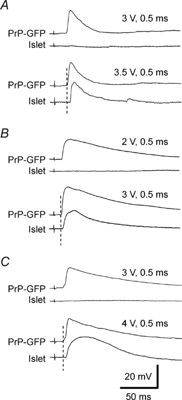
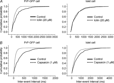
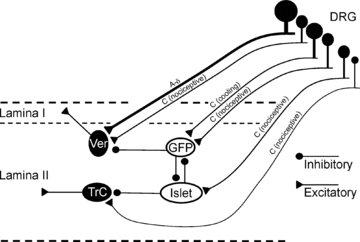
The spinal substantia gelatinosa (SG; lamina II) is a major synaptic zone for unmyelinated (C) primary afferents. Whereas a substantial proportion of intrinsic SG neurones are GABAergic inhibitory, their relationship to afferent activity is unknown. In spinal cord slices from a transgenic mouse in which certain GABAergic lamina II neurones are labelled with green fluorescent protein (GFP), we compared primary afferent input with local efferent connections made by inhibitory SG neurones. Simultaneous whole-cell recordings from characterized neurones establish that inhibitory SG neurones receive monosynaptic input from a subset of unmyelinated primary afferents and connect to other lamina II cells that have input from a different set of afferents, permitting interactions between distinctive afferent messages. Certain lamina II inhibitory cells were found to connect to one another by reciprocal links. Inhibitory lamina II connections appear arranged to modulate activity from different sets of peripheral unmyelinated fibres through neural circuitry that includes disinhibition.
Figures
Similar articles
-
Cell-type-specific excitatory and inhibitory circuits involving primary afferents in the substantia gelatinosa of the rat spinal dorsal horn in vitro.J Physiol. 2007 Jun 1;581(Pt 2):603-18. doi: 10.1113/jphysiol.2006.123919. Epub 2007 Mar 8. J Physiol. 2007. PMID: 17347278 Free PMC article.
-
Primary afferent-evoked glycine- and GABA-mediated IPSPs in substantia gelatinosa neurones in the rat spinal cord in vitro.J Physiol. 1995 Jan 1;482 ( Pt 1)(Pt 1):29-38. doi: 10.1113/jphysiol.1995.sp020497. J Physiol. 1995. PMID: 7730987 Free PMC article.
-
A specific inhibitory pathway between substantia gelatinosa neurons receiving direct C-fiber input.J Neurosci. 2003 Sep 24;23(25):8752-8. doi: 10.1523/JNEUROSCI.23-25-08752.2003. J Neurosci. 2003. PMID: 14507975 Free PMC article.
-
Excitatory interneurons dominate sensory processing in the spinal substantia gelatinosa of rat.J Physiol. 2007 May 15;581(Pt 1):241-54. doi: 10.1113/jphysiol.2006.126912. Epub 2007 Mar 1. J Physiol. 2007. PMID: 17331995 Free PMC article.
-
The role of GABA in primary afferent depolarization.Prog Neurobiol. 1977;9(4):211-67. doi: 10.1016/0301-0082(77)90002-8. Prog Neurobiol. 1977. PMID: 205909 Review. No abstract available.
Cited by
-
Persistent changes in peripheral and spinal nociceptive processing after early tissue injury.Exp Neurol. 2016 Jan;275 Pt 2(0 2):253-60. doi: 10.1016/j.expneurol.2015.06.020. Epub 2015 Jun 21. Exp Neurol. 2016. PMID: 26103453 Free PMC article. Review.
-
Neuronal circuitry for pain processing in the dorsal horn.Nat Rev Neurosci. 2010 Dec;11(12):823-36. doi: 10.1038/nrn2947. Epub 2010 Nov 11. Nat Rev Neurosci. 2010. PMID: 21068766 Free PMC article. Review.
-
Identifying functional populations among the interneurons in laminae I-III of the spinal dorsal horn.Mol Pain. 2017 Jan;13:1744806917693003. doi: 10.1177/1744806917693003. Mol Pain. 2017. PMID: 28326935 Free PMC article. Review.
-
Naked mole-rats lack cold sensitivity before and after nerve injury.Mol Pain. 2020 Jan-Dec;16:1744806920955103. doi: 10.1177/1744806920955103. Mol Pain. 2020. PMID: 32880221 Free PMC article.
-
Excitatory neurons are more disinhibited than inhibitory neurons by chloride dysregulation in the spinal dorsal horn.Elife. 2019 Nov 19;8:e49753. doi: 10.7554/eLife.49753. Elife. 2019. PMID: 31742556 Free PMC article.
References
-
- Bini G, Cruccu G, Hagbarth KE, Schady W, Torebjörk E. Analgesic effect of vibration and cooling on pain induced by intraneural electrical stimulation. Pain. 1984;18:239–248. - PubMed
-
- Burgess PR, Perl ER. Cutaneous mechanoreceptors and nociceptors. In: Iggo A, editor. Handbook of Sensory Physiology, vol II, Somatosensory System. Berlin: Springer-Verlag; 1973. pp. 29–78.
Publication types
MeSH terms
Substances
Grants and funding
LinkOut - more resources
Full Text Sources
Molecular Biology Databases