. 2010 Jun 15;588(Pt 12):2077-90.
doi: 10.1113/jphysiol.2009.185918. Epub 2010 Apr 19.

Unitary inhibitory field potentials in the CA3 region of rat hippocampus

Affiliations

Unitary inhibitory field potentials in the CA3 region of rat hippocampus

Michaël Bazelot et al. J Physiol. .

Abstract

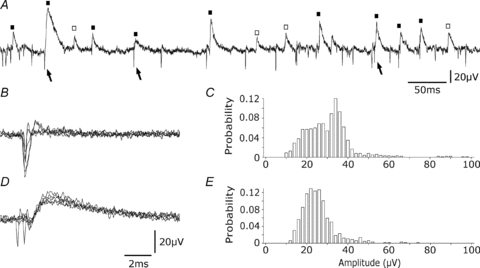
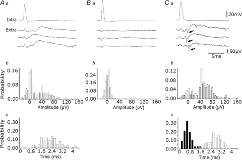
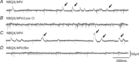
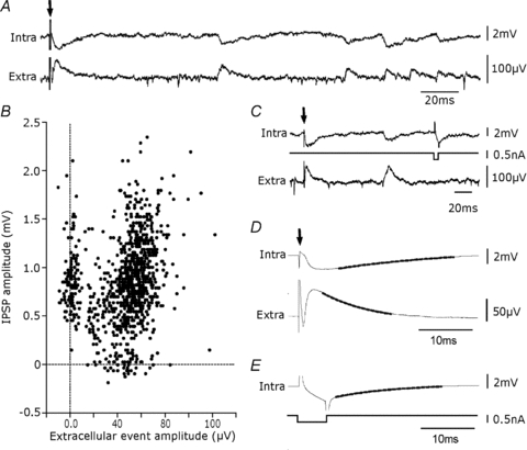
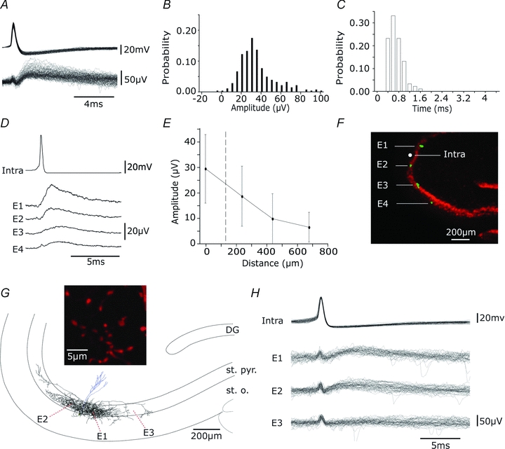
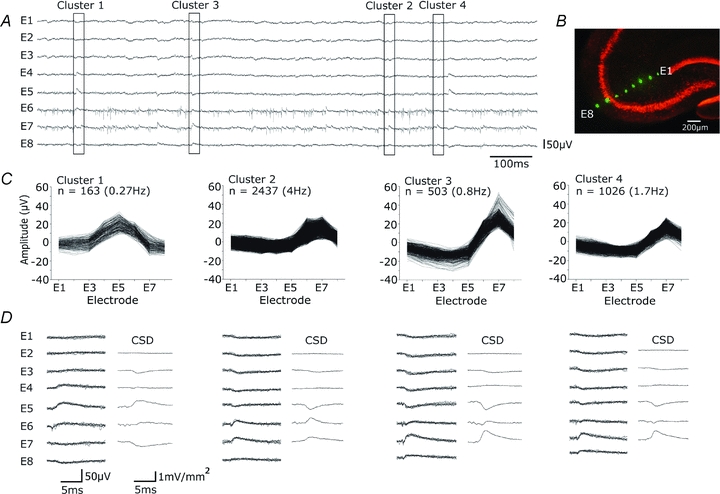
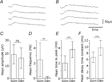
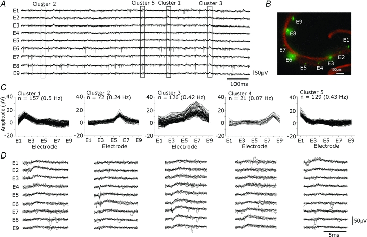
Glickfeld and colleagues (2009) suggested that single hippocampal interneurones generate field potentials at monosynaptic latencies. We pursued this observation in simultaneous intracellular and multiple extracellular records from the CA3 region of rat hippocampal slices. We confirmed that interneurones evoked field potentials at monosynaptic latencies. Pyramidal cells initiated disynaptic inhibitory field potentials, but did not initiate detectable monosynaptic excitatory fields. We confirmed that inhibitory fields were GABAergic in nature and showed they were suppressed at low external Cl(-), suggesting they originate at postsynaptic sites. Field potentials generated by a single interneuron were detected at multiple sites over distances of more than 800 mum along the stratum pyramidale of the CA3 region. We used arrays of extracellular electrodes to examine amplitude distributions of spontaneous inhibitory fields recorded at sites orthogonal to or along the CA3 stratum pyramidale. Cluster analysis of spatially distributed inhibitory field events let us separate events generated by interneurones terminating on distinct zones of somato-dendritic axis. Events generated at dendritic sites had similar amplitudes but occurred less frequently and had somewhat slower kinetics than perisomatic events generated near the stratum pyramidale. In records from multiple sites in the CA3 stratum pyramidale, we distinguished inhibitory fields that seemed to be initiated by interneurones with spatially distinct axonal arborisations.


Figures

Figure 1
Figure 1. Differences between extracellular slow events and action potentials
A, both action potentials (arrows) and slower extracellular events (squares) are evident in an extracellular recording from the stratum pyramidale of the CA3 region. Slow events were sometimes preceded by action potentials (filled squares) and sometimes not preceded (open squares). B, action potentials (5 traces). C, amplitude distribution of 200 successive action potentials. D, slow extracellular events (5 traces). E, amplitude distribution of 200 slow events.
Figure 2
Figure 2. Extracellular events triggered by pyramidal cells
Aa, three slow extracellular events elicited by single action potentials of an intracellularly recorded pyramidal cell. b, amplitude distribution of events (n= 200), measured to their peak or to the peak of the averaged event for signals smaller than 20 μV. About 70% of pyramidal cell spikes evoked an extracellular signal in this recording. c, their latency, from the peak of the intracellular spike to the 10% amplitude of the extracellular event, varied between 1.0 and 4.2 ms. Ba, a pyramidal cell that did not evoke an extracellular event. b, amplitude distribution of extracellular signals (n= 200) measured at 3.5 ms after the peak of the intracellular spike revealed a single peak centred near 0 μV. Ca, interactions initiated by a pyramidal cell, where an extracellular spike (arrow) preceeds each slow event. b, amplitude distribution of extracellular events measured as in Ab (n= 200). c, distribution of the delays between intracellular spikes and extracellular slow events (white bars; n= 100) and between extracellular spikes and slow events (black bars; n= 100). Measurements as in Ac. The peak latency between the extracellular spike and slow event was 0.6 ms and that between the intracellular spike and the slow event was about 3 ms.
Figure 3
Figure 3. Extracellular events are mediated by GABAA receptors
A, extracellular traces showing spikes and extracellular field IPSPs (arrows) recorded in the presence of the glutamate-receptor blockers dl-APV (100 μm) and NBQX (10 μm). B, slow extracellular events were suppressed when the external concentration of Cl was decreased from 130 mm to 50 mm (equimolar replacement by sodium gluconate). Action potential frequency increased. C, slow events reappeared on return to the original Cl concentration. D, the GABAA receptor antagonist bicuculline (20 μm) suppressed slow extracellular events.
Figure 4
Figure 4. Intracellular correlates of extracellular events
A, intracellular correlates (intra) of extracellular events (extra) occurring spontaneously and elicited by weak local stimulation (stimulation, arrow, in stratum pyramidale at 200 μm from the recorded pyramidal cell, extracellular electrode separated by ∼100 μm from the pyramidal cell). Many events were correlated. Records made in the presence of dl-APV (100 μm) and NBQX (10 μm). B, amplitude of spontaneous inhibitory field events plotted against that of intracellular IPSPs. Measurements were made to the peak of respective events when either event was detected (n= 988). In this recording about 75% (n= 727) of intracellular IPSPs were accompanied by an extracellular signal with a difference in onset times of less than 2 ms. Their correlation coefficient, r, was 0.46 (r2= 0.21, P < 0.0001, Pearson). About 9% of extracellular signals were not accompanied by an intracellular IPSP and 16% of IPSPs had no extracellular correlate. C, comparison of decay kinetics for the evoked IPSP and response to a small intracellular current injection and the evoked extracellular field. D, the intracellular IPSP decayed with a time constant of 23.9 ± 0.1 ms and the field event with a time constant of 9.8 ± 0.4 ms. E, the response to a 5 ms hyperpolarizing current step in the same cell decayed with a time constant of 18.3 ± 0.2 ms. Traces in D and E are averages (n= 300) with a mono-exponential fit shown as a thicker line.
Figure 5
Figure 5. Extracellular events induced by interneurone firing are detected at multiple sites along stratum pyramidale
A, overlay of 100 events elicited by single action potentials of an interneurone. B, distribution of amplitudes (n= 300). C, distribution of latencies (n= 100). D, averages (n= 50) of extracellular events, initiated by single spikes in an interneurone (intra), recorded by 4 extracellular electrodes. Electrodes, E1–E4, were located in CA3 stratum pyramidale at separations of about 200 μm. E, mean and standard deviations of the amplitude of extracellular events (n= 50) plotted against distance from the recording site. F, localization of recording sites with DiO staining. Electrodes were coated with DiO (green) before recording and neuronal somata are shown by immunostaining for NeuN (red). G, reconstruction of the dendritic (blue) and axonal (black) arbour of a biocytin-filled perisomatic cell. Axon terminals were largely confined to the CA3 stratum pyramidale (st. pyr.) with some in stratum oriens (st. o.), and were distributed over about 1mm along CA3 st. pyr. The inset shows axon terminals of diameter 1–2 μm. Extracellular records were made from sites E1–3 indicated by red dots. H, single action potentials evoked field IPSPs in two of three extracellular records. Thirty responses from electrodes E1–E3 are shown.
Figure 6
Figure 6. Extracellular signals recorded along the CA3 pyramidal cell somato-dendritic axis
A, spontaneous extracellular signals recorded from multiple sites orthogonal to the CA3 stratum pyramidale with 8 electrodes separated by ∼100 μm, in the presence of dl-APV (100 μm) and NBQX (10 μm). Extracellular spikes were limited to electrodes E6 and E7, while field IPSPs were evident in signals from E1–E8. B, extracellular recording sites, marked with DiO (green), and neuronal somata by NeuN immunostaining (red). C and D, K-means cluster analysis separated distinct patterns of signal from the 8 electrodes for 4129 events recorded during 10 min (6.8 Hz). C, amplitude distributions for 8 signals and event frequencies from 4 clusters. Cluster 1 corresponds to an infrequent event with maximal amplitude in stratum radiatum, while clusters 2–4 show more frequent events with different distributions and amplitude maxima around stratum pyramidale. D, records from each cluster. At left, five superimposed traces from each electrode (E1–E8). At right, current source density analysis as the discrete second spatial derivative of averaged extracellular fields.
Figure 7
Figure 7. Differences between perisomatic and dendritic inhibitory fields
Dendritic and perisomatically generated fields were separated by current source density analysis of clusters from 6 electrodes. A, perisomatic field IPSPs of maximal amplitude in stratum pyramidale. B, dendritic field IPSPs of maximal amplitude from an electrode in CA3 stratum radiatum. C–F, comparison of amplitude, frequency, time to peak and decay for perisomatic (Som) and dendritic (Den) field events. C, the amplitude of perisomatic and dendritic field IPSPs was not different (Som, 19.1 ± 7.6 μV, n= 18 clusters; Den, 16.2 ± 8 μV, n= 10 clusters; ns, P= 0.34, t test). D, the frequency of events from dendritic field IPSP clusters was lower than that of perisomatic events (Som, 2.45 ± 2.2 Hz, n= 18 clusters; Den, 0.17 ± 0.12 Hz, n= 10 clusters; **P < 0.001, t test). E, the time to peak, measured from 10 to 90% peak amplitude, was significantly longer for dendritic field IPSPs (Som, 2.0 ± 0.9 ms, n= 18 clusters; Den, 3.8 ± 1 ms, n= 10 clusters; ***P < 0.0001, t test). F, the mean decay time constant (mono-exponential decay fitted to 10–90% amplitude decay for averaged events) was significantly slower for dendritic field IPSPs events (Som, 6.6 ± 1.6 ms, n= 18 clusters; Den, 9.6 ± 2.3 ms, n= 10 clusters; ***P < 0.0005, t test).
Figure 8
Figure 8. Spontaneous inhibitory field distributions along the CA3 stratum pyramidale
A, extracellular signals recorded from multiple sites of the CA3 stratum pyramidale with 9 extracellular electrodes separated by ∼250 μm. Records made in the presence of dl-APV (100 μm) and NBQX (10 μm). Extracellular spikes, evident on all electrodes, were rarely simultaneous. Field IPSPs, of differing forms and amplitudes were evident at all sites and often occurred simultaneously at several neighbouring electrodes. B, location of extracellular recording sites, marked with DiO (green), and neuronal cell bodies detected by NeuN immunostaining for (red). C and D, K-means cluster analysis was done to separate distinct signal patterns. For 501 events recorded over 5 min, K-means clustering was performed on amplitude values from all electrodes. C, shows amplitude distributions from 9 electrodes and the frequencies of events of five clusters in this recording. Each cluster seems to have a distinct at different regions of the CA3 stratum pyramidale. D, an overlay of five traces from each electrode (E1–E9) for each cluster.

Comment in

Similar articles

Cited by

References

    1. Andersen P, Bliss TV, Skrede KK. Unit analysis of hippocampal population spikes. Exp Brain Res. 1971;13:208–221. - PubMed
    1. Banks MI, Li TB, Pearce RA. The synaptic basis of GABAA,slow. J Neurosci. 1998;18:1305–1317. - PMC - PubMed
    1. Bliss TV, Lomo T. Long-lasting potentiation of synaptic transmission in the dentate area of the anaesthetized rabbit following stimulation of the perforant path. J Physiol. 1973;232:331–335. - PMC - PubMed
    1. Brecht M, Schneider M, Sakmann B, Margrie TW. Whisker movements evoked by stimulation of single pyramidal cells in rat motor cortex. Nature. 2004;427:704–710. - PubMed
    1. Biernacki C, Celeux G, Govaert G, Langrognet F. Model-based cluster and discriminant analysis with the MIXMOD software. Comput Stat Data Anal. 2006;51(2):587–600.

Publication types

MeSH terms

Substances