. 2010 Jul;78(7):2966-73.
doi: 10.1128/IAI.01284-09. Epub 2010 Apr 19.

Rapid cytopathic effects of Clostridium perfringens beta-toxin on porcine endothelial cells

Affiliations

Rapid cytopathic effects of Clostridium perfringens beta-toxin on porcine endothelial cells

Corinne Gurtner et al. Infect Immun. 2010 Jul.

Abstract

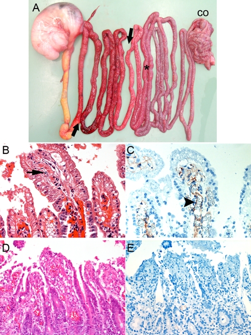
Clostridium perfringens type C isolates cause fatal, segmental necro-hemorrhagic enteritis in animals and humans. Typically, acute intestinal lesions result from extensive mucosal necrosis and hemorrhage in the proximal jejunum. These lesions are frequently accompanied by microvascular thrombosis in affected intestinal segments. In previous studies we demonstrated that there is endothelial localization of C. perfringens type C beta-toxin (CPB) in acute lesions of necrotizing enteritis. This led us to hypothesize that CPB contributes to vascular necrosis by directly damaging endothelial cells. By performing additional immunohistochemical studies using spontaneously diseased piglets, we confirmed that CPB binds to the endothelial lining of vessels showing early signs of thrombosis. To investigate whether CPB can disrupt the endothelium, we exposed primary porcine aortic endothelial cells to C. perfringens type C culture supernatants and recombinant CPB. Both treatments rapidly induced disruption of the actin cytoskeleton, cell border retraction, and cell shrinkage, leading to destruction of the endothelial monolayer in vitro. These effects were followed by cell death. Cytopathic and cytotoxic effects were inhibited by neutralization of CPB. Taken together, our results suggest that CPB-induced disruption of endothelial cells may contribute to the pathogenesis of C. perfringens type C enteritis.


Figures

FIG. 1.
FIG. 1.
Pathology and immunohistochemistry. (A) Distribution of macroscopic lesions of acute C. perfringens type C enteritis in a 2-day-old piglet (stomach on left side; CO, colon). The arrows indicate the extent of typical necro-hemorrhagic lesions in the proximal jejunum. The asterisk indicates a histopathological sampling site in a macroscopically unaffected segment of the caudal jejunum. (B) H&E-stained histological sections of a macroscopically unaffected segment, showing early signs of epithelial degeneration at villous tips and thrombus formation (arrow) in an underlying vessel. (C) Immunohistochemical labeling with MAb-CPB of a serial section of the specimen shown in panel B, showing positive staining on the endothelial lining of a vessel (arrowhead). Note that no immunoreactivity was observed with epithelial cells. (D) H&E-stained control tissue from a piglet with superficial necrotizing enteritis due to I. suis infection. (E) Immunohistochemistry of sections revealed no CPB specific staining. (B and C) Magnification, ×1,000. (D and E) Magnification, ×400.
FIG. 2.
FIG. 2.
Cytotoxicity of C. perfringens type C culture supernatants for PAEC. (A) MTT cell viability tests using serial dilutions of culture supernatants revealed that JF 3719 and JF 3721 (type C), but not JF 3693 or JF 3686 (type A), induced dose-dependent cytotoxicity in PAEC. (B) Preincubation of JF 3719 and JF 3721 supernatants (1:20 [vol/vol] dilutions; 125 ng/ml CPB) with different amounts of MAb-CPB inhibited cytotoxicity for PAEC in a dose-dependent manner. Preincubation with MAb-CPA did not have an inhibitory effect. The exposure time was 5 h. MTT values were obtained in duplicate for each time point. The symbols indicate the means for two separate experiments performed with PAEC from one donor. The error bars indicate the standard errors of the means. Asterisks indicate significant differences between type C supernatants and control type A supernatants (A) or between preincubation with MAb-CPB and preincubation with MAb-CPA (B) (P < 0.05).
FIG. 3.
FIG. 3.
Expression of rCPB: Western blot analysis of 10-μl aliquots of purified rCPB-Nus-Tag (expected molecular mass, 98 kDa) (lane 1), rCPB (expected molecular mass, 35 kDa) after thrombin cleavage (lane 2), and rCPB after thrombin elimination (lane 3). Western blots were developed with MAb-CPB. Additional bands in lanes 2 and 3 represent degradation products of the fusion protein, likely due to excessive thrombin activity.
FIG. 4.
FIG. 4.
Cytotoxicity of rCPB for PAEC. (A) Exposure of PAEC to rCPB, but not exposure of PAEC to rCPB-Nus-Tag or mock-purified Nus-Tag, resulted in a dose-dependent reduction in cell viability. (B) Preincubation of rCPB with MAb-CPB inhibited cytotoxicity for PAEC in a dose-dependent manner. The exposure time was 12 h. MTT values were obtained from two separate experiments performed in duplicate for each time point. The symbols indicate the means for two separate experiments with PAEC from one donor. The error bars indicate the standard errors of the means. The asterisks in panel A indicate statistical significance for comparisons of rCPB with both control preparations (P < 0.05). The asterisks in panel B indicate statistical significance for comparisons of rCPB with 1,000- and 10,000-fold dilutions of MAb-CPB and control preparations (P < 0.05).
FIG. 5.
FIG. 5.
CPB-induced cytopathic effects on PAEC: photomicrographs of PAEC after 3 h of exposure to serum-free medium (SFM), a JF 3719 culture supernatant dilution containing 32 ng/ml CPB, 32 ng/ml rCPB, and a 1:10 (vol/vol) dilution of JF 3693 culture supernatant. Only CPB-containing media induced cell shrinkage and widening of intercellular gaps. Preincubation with neutralizing anti-CPB monoclonal antibodies (mAb-CPB) inhibited the cytopathic effects. Cells were fixed in methanol and stained with Diff-Quik. Magnification, ×400.
FIG. 6.
FIG. 6.
rCPB-induced disruption of the actin cytoskeleton in PAEC: fluorescent staining of F-actin in PAEC after 1 and 3 h and in porcine fibroblasts after 3 h of incubation with serum-free medium (SFM), 32 ng/ml rCPB, 32 ng/ml rCPB preincubated with MAb-CPB, and 3 μg/ml rCPB-Nus-Tag fusion protein. Disruption of the actin cytoskeleton was visible 1 h after exposure to rCPB. The effect increased over time (3 h) and was inhibited by neutralization of rCPB. The actin cytoskeleton of porcine fibroblasts was not affected.

References

    1. Albini, S., I. Brodard, A. Jaussi, N. Wollschlaeger, J. Frey, R. Miserez, and C. Abril. 2008. Real-time multiplex PCR assays for reliable detection of Clostridium perfringens toxin genes in animal isolates. Vet. Microbiol. 127:179-185. - PubMed
    1. Amimoto, K., T. Noro, E. Oishi, and M. Shimizu. 2007. A novel toxin homologous to large clostridial cytotoxins found in culture supernatant of Clostridium perfringens type C. Microbiology 153:1198-1206. - PubMed
    1. Awad, M. M., D. M. Ellemor, R. L. Boyd, J. J. Emmins, and J. I. Rood. 2001. Synergistic effects of alpha-toxin and perfringolysin O in Clostridium perfringens-mediated gas gangrene. Infect. Immun. 69:7904-7910. - PMC - PubMed
    1. Bouis, D., G. A. Hospers, C. Meijer, G. Molema, and N. H. Mulder. 2001. Endothelium in vitro: a review of human vascular endothelial cell lines for blood vessel-related research. Angiogenesis 4:91-102. - PubMed
    1. Bryant, A. E. 2003. Biology and pathogenesis of thrombosis and procoagulant activity in invasive infections caused by group A streptococci and Clostridium perfringens. Clin. Microbiol. Rev. 16:451-462. - PMC - PubMed

Publication types

MeSH terms

Substances

LinkOut - more resources