. 2010 Jul;30(13):3187-205.
doi: 10.1128/MCB.00227-10. Epub 2010 Apr 19.

Two-step imprinted X inactivation: repeat versus genic silencing in the mouse

Affiliations

Two-step imprinted X inactivation: repeat versus genic silencing in the mouse

Satoshi H Namekawa et al. Mol Cell Biol. 2010 Jul.

Abstract

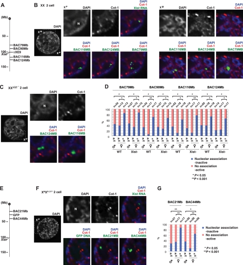
Mammals compensate for unequal X-linked gene dosages between the sexes by inactivating one X chromosome in the female. In marsupials and in the early mouse embryo, X chromosome inactivation (XCI) is imprinted to occur selectively on the paternal X chromosome (X(P)). The mechanisms and events underlying X(P) imprinting remain unclear. Here, we find that the imprinted X(P) can be functionally divided into two domains, one comprising traditional coding genes (genic) and the other comprising intergenic repetitive elements. X(P) repetitive element silencing occurs by the two-cell stage, does not require Xist, and occurs several divisions prior to genic silencing. In contrast, genic silencing initiates at the morula-to-blastocyst stage and absolutely requires Xist. Genes translocate into the presilenced repeat region as they are inactivated, whereas active genes remain outside. Thus, during the gamete-embryo transition, imprinted XCI occurs in two steps, with repeat silencing preceding genic inactivation. Nucleolar association may underlie the epigenetic asymmetry of X(P) and X(M). We hypothesize that transgenerational information (the imprint) is carried by repeats from the paternal germ line or that, alternatively, repetitive elements are silenced at the two-cell stage in a parent-of-origin-specific manner. Our model incorporates aspects of the so-called classical, de novo, and preinactivation hypotheses and suggests that Xist RNA functions relatively late during preimplantation mouse development.


Figures

FIG. 1.
FIG. 1.
Repeat silencing of XP in the early embryo. (A to C) Two-color RNA FISH reveals the relationship between Xist RNA and Cot-1 expression in three representative XX two-cell embryos, using published protocols (47) (A and B; both nuclei are shown in the right panels) or our newly developed protocols (C). All images are deconvolved single z sections. Blastomeres indicated by asterisks in leftmost panels are magnified. Results for the second blastomere are shown in Fig. S1 in the supplemental material. The intensity of Cot-1 expression is quantified by fluorimetry across the indicated path (′ to ″) and plotted in the relative intensity range of 0 to 1. Note that XP often resides next to the prenucleolus (N) and coincides with DAPI-intense regions. (D) X chromosome painting of a two-cell blastomere reveals that XP and XM occupy large nuclear territories. Note that XP is associated with the prenucleolus and is relatively lacking in Cot-1 compared to XM. The image represents merged z stacks (3D stacks projected onto a single plane) to capture X-chromosomal signals in multiple z sections. (E to H) Cot-1 RNA FISH of later stage XX embryos as indicated, presented as described for panel A to C. For panel G and H, serial z sections are merged (merged z's) to show the degree of Cot-1 expression throughout the nucleus. Following slide denaturation, X paint was performed to reveal the locations of XM and XP.
FIG. 2.
FIG. 2.
Characterization of the Cot-1 domain. A single representative blastomere is shown in all panels, and the intensity of immunostaining is quantified by densitometry across the indicated path (′ to ″) and plotted in the relative intensity range of 0 to 1. (A to C) Immunostaining using antibodies against RNA polymerases II and III as indicated. The frequencies of exclusion from XP were the following: Pol-II (CTD), 59% (n = 17); Pol-II (H5), 79% (n = 19); Pol-III, 58% (n = 19). (D to F) Expression analysis of the indicated repetitive elements by RNA FISH. Exclusion frequencies were the following: LINE, 62% (n = 13); B1, 67% (n = 12); and B2, 64% (n = 14).
FIG. 3.
FIG. 3.
Genic expression of XP and XM in the early embryo. (A) RNA/DNA FISH to confirm the specificity of RNA FISH signals. To ensure that nascent RNA signals originated from the corresponding genes, images were captured, cell coordinates marked, and slides denatured for subsequent DNA FISH using the same BAC probe (labeled with a different fluorophore). Note that RNA and DNA signals were perfectly coincident, confirming the specificity of RNA FISH. A representative experiment is shown. (B, D, G, and I) Nascent RNA FISH of indicated genes combined with Xist RNA FISH. Representative nuclei are shown. For all images, z sections were taken through the nucleus and merged into one plane to view all signals. (C, F, H, and J) Summary of all RNA FISH data from female (F) and male (M) embryos of the indicated stage. n, number of nuclei examined; the number of embryos analyzed is in parentheses. (E) Example of skewed expression in which the XP RNA shows fewer nascent transcripts at the four-cell stage.
FIG. 4.
FIG. 4.
Translocation of genic loci into the silent repeat compartment during silencing. (A) Active XP genes reside outside of the Xist+ Cot-1 repeat compartment before silencing. Three representative blastomeres from morulae are shown expressing three X-linked genes. Arrows indicate linear distances between the gene and the outer edge of the Xist+ Cot-1 compartment. (B) During the process of silencing (deduced by diminished XP expression), genes are translocated into the Xist+ Cot-1 compartment in the morula. The boxed region is magnified in the right panel. Arrows indicate linear distances between the gene and the outer edge of the Xist+ Cot-1 compartment. (C) Summary of linear distances between the gene and the silent compartment during silencing. The normalized distance is the linear distance from the center of the nascent RNA signal to the edge of the Xist+ Cot-1 compartment, each normalized to the nuclear diameter. Negative distances imply genic movement into the Xist+ Cot-1 compartment, whereas a zero distance implies localization at the edge of the Xist+ Cot-1 compartment. P values were calculated using an unpaired t test. (D) XP territory contracts over time. XP and XM territories were measured by Volocity software (Improvision) and normalized to total nuclear volume to yield the chromosome condensation index. P values were calculated using an unpaired t test. (E) Pictorial representation of genic localization into the preformed Xist+ Cot-1 compartment during silencing. The silent compartment is present by the two-cell stage, and it enlarges as genic loci are translocated into it as they are gradually inactivated, beginning at the morula stage. XP silencing is not complete until the blastocyst stage or later.
FIG. 5.
FIG. 5.
Genic silencing depends on Xist. (A to D) Biallelic expression of indicated genes in Xist-deficient preimplantation embryos of different stages. Shown are merged z sections taken through the nucleus to capture signals in all focal planes. (E to H) Summary of genic expression in Xist mutant female (F) and male (M) embryos of the indicated stage. n, number of nuclei examined; the number of embryos analyzed is in parentheses. (I) Allele-specific RT-PCR of seven X-linked genes in wild-type (WT) and Xist mutant morulae produced by the indicated crosses. Mus, M. musculus; Cas, M. castaneus; M, maternal; P, paternal; PBS, negative control derived from the wash fluid after embryos are isolated to rule out contamination.
FIG. 6.
FIG. 6.
Analysis of imprinted XCI using an X-linked GFP transgene. (A to D) Blastocysts isolated at E3.5 (A and B) or cultured for one additional day to E4.5. (C and D) The embryos carry a paternally transmitted X-linked GFP transgene on either a wild-type X (A and C) or Xist-deficient X (B and D). GFP expression is evident even when GFP is carried on a wild-type XP, consistently with a later onset of genic XP silencing than that expected and consistently with the produrance of GFP. Bright-field (left) and GFP fluorescence (right) images are shown for each embryo. (C) Male embryos without XP are nontrangenic for X-linked GFP and are therefore GFP−. ICM, inner cell mass; TE, trophectoderm. (E to H) Embryo outgrowths attached as blastocysts and cultured for 2 to 3 days until E5.5 (E and F) or E6.5 (G and H). Each embryo carries the paternally transmitted X-linked GFP transgene on a wild-type X (E and G) or Xist-deficient X (F and H). epi, epibast; ExE, extraembryonic ectoderm. (I) Quantitative assessment of X-linked GFP fluorescence for the stages shown in panels A to H. The mean fluorescence intensity was measured for each stage in the embryonic parts (ICM or epi) and in the extraembryonic parts (TE or ExE) as depicted in panels A and E. While no significant GFP signal differences can be quantified in E3.5 and E4.5 blastocysts, later on wild-type embryos show lower GFP signals than Xist-deficient embryos, which is consistent with the onset of genic XP silencing. Fluorescence differences between wild-type and Xist mutant embryos are statistically significant at E6.5 according to the nonpaired t test (epi, P = 0.0015; ExE, P = 0.0314). Error bars indicate standard errors of the means. (J to O) E6.5 embryos of the indicated genotypes as dissected from maternal deciduas (K to O) and the quantitation of the GFP signals depicted in panel L (J). GFP is almost silent in ExE regardless of whether it is carried on a wild-type or Xist mutant X chromosome and whether it is paternally or maternally transmitted, despite the fact that there is no imprinted inactivation of the maternal X in female (J, M, and N) or male (J and O) embryos. Therefore, the GFP transgene cannot be used as a reliable marker to analyze imprinted XCI. het, the mother is heterozygous for X-GFP; hom, the mother is homozygous for X-GFP.
FIG. 7.
FIG. 7.
Repeat silencing in the early preimplantation embryo does not require Xist. Cot-1 and Xist RNA FISH with subsequent DNA FISH using the Xic probe, Sx7, in the wild-type two-cell embryo (A), and Cot-1 RNA FISH with subsequent DNA FISH in the Xist mutant two-cell embryo using a combination of Sx7 and πXE9 probes to distinguish XM (wild-type) from XP (Xist deficient) (B). (C) Pictorial representation of the DAPI staining pattern and corresponding Cot-1 RNA FISH pattern at the two-cell stage. Because the nucleolus and perinucleolar heterochromatin are devoid of Cot-1 signal, the Cot-1 hole is always larger than the DAPI hole left by the nucleolus itself. When the DNA FISH signal in question localizes to the perinucleolar Cot-1 hole, the signal is scored as nucleolar association inactive. On the other hand, when the signal localizes in Cot-1+ regions, it is scored as no association active. (D) Xic nucleolar association of XP versus XM. Cot-1, LINE, B1, and B2 RNA FISH were performed on wild-type and Xist-deficient two-cell embryos in combination with an Xic probe (Sx7). DNA FISH was conducted subsequently to compare the frequency with which XP and XM come in direct contact with the nucleolus. The combination of Sx7 and πXE9 probes was used to distinguish XM (wild-type) from XP (Xist deficient). When the Sx7 signal was directly adjacent to the nucleolus or located in the perinucleolar heterochromatic ring, the chromosome was judged to be nucleolus associated and Cot-1. For the two-cell embryo, there was 100% correlation between nucleolus-associated and Cot-1, LINE, and SINE B2 states of XP; there was a 95% correlation between the nucleolus-associated and the SINE B1 state. In contrast, chromosomes that were not nucleolus associated were Cot-1+. P values were calculated using the student t test. WT, wild-type. Xist−, XMXP;Xist.
FIG. 8.
FIG. 8.
Repetitive elements outside of the Xist RNA domain on XP also are silenced in an Xist-independent manner (A) Locations of repeat-rich X-linked BAC sequences examined in panels B and C. Each region is located outside of the Xist RNA-coated domain of XP in the two-cell embryo. (B and C) Cot-1 and Xist RNA FISH with subsequent DNA FISH of repetitive elements outside of the Xist RNA-coated domain in the wild-type two-cell embryo (B) or the XMXP;Xist two-cell embryo (C). One z section is shown. Note that some BAC probes yielded multiple signals, possibly reflecting sister chromatids of a cell in the G2 stage of the cell cycle and/or the longer probe lengths of some BACs (200 to 300 kb), which would yield a linear track of signals. (D) Quantitation of nucleolar association and repeat silencing for the experiments shown in panel B and C. Cot-1 RNA FISH was performed on wild-type and Xist-deficient embryos as described for Fig. 7D. P values were calculated using the χ2 test. (E) Locations of repeat-rich X-linked BAC probes for the analysis shown in panels F and G. (F) Cot-1 and Xist RNA FISH with subsequent DNA FISH in the wild-type two-cell embryo with a paternally transmitted GFP transgene. One z section is shown. (G) Quantitation of nucleolar association and repeat silencing for the experiments shown in panel F. P values were calculated using the unpaired student t test.
FIG. 9.
FIG. 9.
Perinucleolar association present in two- and four-cell embryos is lost during the 8-cell stage. (A) Cot-1 and Xist RNA FISH with subsequent DNA FISH using the Xic probe, Sx7, in the wild-type four-cell embryo. (B) Cot-1 RNA FISH with subsequent DNA FISH in the Xist mutant four-cell embryo using a combination of Sx7 and πXE9 probes to distinguish XM (wild-type) from XP (Xist deficient). For panels A and B, two z sections (top and bottom) are shown for each blastomere to capture the XM and XP planes. (C) Xic nucleolar association of XP versus that of XM in 4- and 8-cell embryos. Cot-1 RNA FISH was performed on wild-type and Xist-deficient embryos as described for Fig. 7C with the following exception: for 4- and 8-cell embryos, nucleolar association correlated with a Cot-1 state of XP in 100% of blastomeres; however, for the wild-type embryos, a lack of nucleolar association was correlated with silencing in a fraction of blastomeres. P values were calculated using the unpaired student t test. WT, wild-type. Xist−, XMXP;Xist. (D) X chromosome painting of a two-cell embryo of an Xist mutant reveals large X territories (circled) at this stage. (E) Cot-1 RNA FISH and subsequent X chromosome painting show no compaction of XP in the Xist mutant embryo at the blastocyst stage. (F) Pictorial representation of the deduced XP and XM structures in the early embryo. Repeat elements of XP lie in the silent perinucleolar compartment, while XM and active genic loci of XP reside in Cot-1+ regions.
FIG. 10.
FIG. 10.
Postmeiotic silencing also does not require Xist. (A and B) Cot-1 RNA FISH and the chromosome-specific painting of X and Y in the round spermatids of wild-type mice. Arrow, PMSC. Single z sections are shown for all panels. (C and D) Immunofluorescence for HP1β (C) and H3-2meK9 (D) in wild-type round spermatids. (E and F) Cot-1 RNA FISH and chromosome-specific painting of X and Y in the round spermatids of Xist-deficient mice. (G and H) Immunofluorescence for HP1β (G) and H3-2meK9 (H) in mutant round spermatids. (I) A working hypothesis for the developmental history of the X chromosome from gamete to embryo.

References

    1. Adler, D., J. West, and V. Chapman. 1977. Expression of alpha-galactosidase in preimplantation mouse embryos. Nature 267:838-839. - PubMed
    1. Avner, P., and E. Heard. 2001. X-chromosome inactivation: counting, choice and initiation. Nat. Rev. Genet. 2:59-67. - PubMed
    1. Bartolomei, M. S., and S. M. Tilghman. 1997. Genomic imprinting in mammals. Annu. Rev. Genet. 81:493-525. - PubMed
    1. Bean, C. J., C. E. Schaner, and W. G. Kelly. 2004. Meiotic pairing and imprinted X chromatin assembly in Caenorhabditis elegans. Nat. Genet. 36:100-105. - PMC - PubMed
    1. Beatty, B., S. Mai, and J. Squire. 2002. FISH. A practical approach. Oxford University Press, Oxford, United Kingdom.

Publication types

LinkOut - more resources