. 2010 Jun;30(12):3071-85.
doi: 10.1128/MCB.01428-09. Epub 2010 Apr 19.

Osteocyte Wnt/beta-catenin signaling is required for normal bone homeostasis

Affiliations

Osteocyte Wnt/beta-catenin signaling is required for normal bone homeostasis

Ina Kramer et al. Mol Cell Biol. 2010 Jun.

Abstract

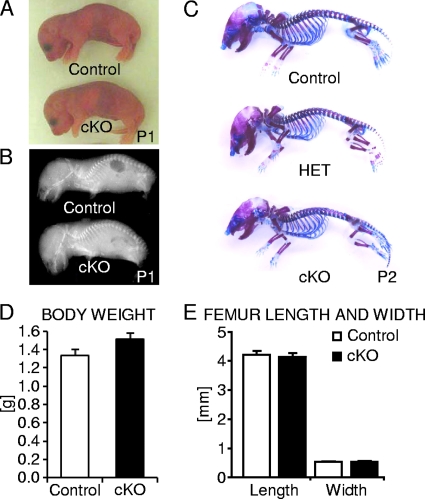
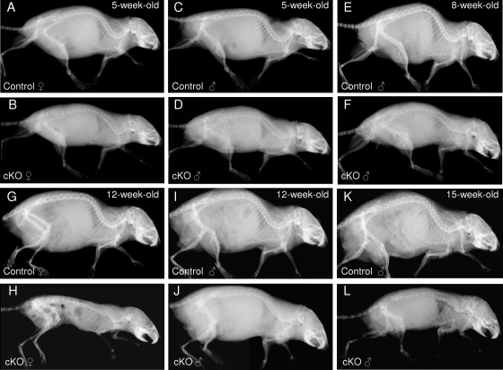
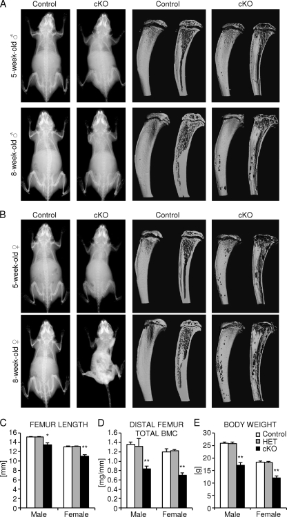
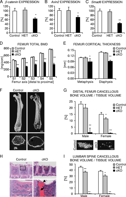
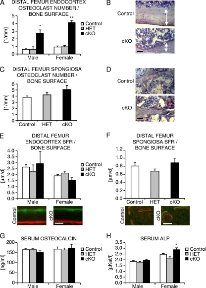
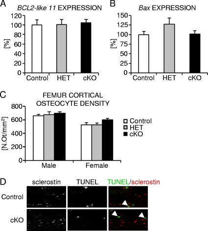
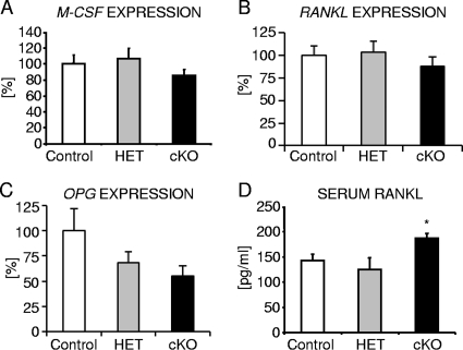
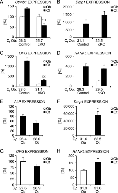
Beta-Catenin-dependent canonical Wnt signaling plays an important role in bone metabolism by controlling differentiation of bone-forming osteoblasts and bone-resorbing osteoclasts. To investigate its function in osteocytes, the cell type constituting the majority of bone cells, we generated osteocyte-specific beta-catenin-deficient mice (Ctnnb1(loxP/loxP); Dmp1-Cre). Homozygous mutants were born at normal Mendelian frequency with no obvious morphological abnormalities or detectable differences in size or body weight, but bone mass accrual was strongly impaired due to early-onset, progressive bone loss in the appendicular and axial skeleton with mild growth retardation and premature lethality. Cancellous bone mass was almost completely absent, and cortical bone thickness was dramatically reduced. The low-bone-mass phenotype was associated with increased osteoclast number and activity, whereas osteoblast function and osteocyte density were normal. Cortical bone Wnt/beta-catenin target gene expression was reduced, and of the known regulators of osteoclast differentiation, osteoprotegerin (OPG) expression was significantly downregulated in osteocyte bone fractions of mutant mice. Moreover, the OPG levels expressed by osteocytes were higher than or comparable to the levels expressed by osteoblasts during skeletal growth and at maturity, suggesting that the reduction in osteocytic OPG and the concomitant increase in osteocytic RANKL/OPG ratio contribute to the increased number of osteoclasts and resorption in osteocyte-specific beta-catenin mutants. Together, these results reveal a crucial novel function for osteocyte beta-catenin signaling in controlling bone homeostasis.


Figures

FIG. 1.
FIG. 1.
Analysis of newborn osteocyte-specific β-catenin gene cKO mice. (A and B) Representative photographic images (A) and whole-body skeletal radiographies (B) of postnatal day 1 (P1) homozygous osteocyte-specific β-catenin-deficient (cKO) and control mice. (C) Whole-mount skeleton staining of P2 homozygous (cKO) and heterozygous (HET) osteocyte-specific β-catenin cKO and control mice. (D and E) Quantification of body weight (D) and femoral length and width (E) of P1 cKO and control littermates. The values are means plus standard errors of the means (error bars). There were six mice in each group.
FIG. 2.
FIG. 2.
Time course of the skeletal phenotype of osteocyte-specific β-catenin cKO mice. Representative whole-body skeletal radiographies of 5-week-old (A to D), 8-week-old (E and F), 12-week-old (G to J), and 15-week-old (K and L) homozygous osteocyte-specific β-catenin-deficient (cKO) and control littermate female (A, B, G, and H) and male (C to F and I to L) mice.
FIG. 3.
FIG. 3.
Analysis of juvenile osteocyte-specific β-catenin cKO mice. (A and B) Representative whole-body skeletal radiographies (left two columns) and μCT images of the proximal tibia to the midshaft level (right two columns) of 1-month-old (top row) and 2-month-old (bottom row) homozygous osteocyte-specific β-catenin-deficient (cKO) and control littermate male mice (A) and female mice (B). (C to E) Quantification of femoral length (C), cross-sectional total BMC in the distal femur metaphysis (D), and body weight (E) of 2-month-old homozygous (cKO) and heterozygous (HET) osteocyte-specific β-catenin-deficient and control littermates. There were 3 to 11 mice in a group. Values for the mutant that were significantly different from the value for control littermate mice of the same gender using unpaired Student's t tests are shown as follows: *, P < 0.05; **, P < 0.01.
FIG. 4.
FIG. 4.
Characterization of femoral and vertebral bone of osteocyte-specific β-catenin cKO mice. (A to C) Relative expression normalized to 18S for β-catenin (A) and downstream Wnt/β-catenin target genes Axin2 (B) and Smad6 (C) in cortical femoral bone from 2-month-old female mice. (D and E) Femoral cross-sectional total BMD (D) and cortical thickness (E) of 2-month-old homozygous (cKO) and heterozygous (HET) β-catenin-deficient and control littermate female mice as evaluated by ex vivo pQCT analyses (D) of five consecutive slices S1 to S5 spaced equally along the femoral axis from the distal metaphysis (S1) to the proximal end of the diaphysis (S5) or by ex vivo μCT analyses (E) in the distal metaphysis and diaphysis. (F) Representative μCT images of the femur from 2-month-old homozygous β-catenin-deficient (cKO) and control littermate female mice. (G and I) Quantification of relative cancellous bone volume in the distal femur metaphysis with representative 3D-reconstructed μCT images from male mice (G) and lumbar vertebra L3 (I) of 2-month-old homozygous (cKO) and heterozygous (HET) β-catenin-deficient and control littermates. (H) Representative H&E stained images of thoracic vertebrae from 3-month-old homozygous cKO and control female mice depicting a vertebral fracture (arrowhead) and extensive scar tissue formation (arrow). The boxes outlined by a broken line in the top row are shown at higher magnification in the bottom row. There were 3 to 11 mice in each group. Values for the mutant that were significantly different from the value for control littermate mice using unpaired Student's t tests are shown as follows: *, P < 0.05; **, P < 0.01. Bars, 1 mm (F, top row), 0.5 mm (F, bottom row), 0.5 mm (G), 0.25 mm (H, magnified view).
FIG. 5.
FIG. 5.
Histomorphometric analysis of femoral bone of osteocyte-specific β-catenin cKO mice. (A, C, E, and F) Quantification of the number of osteoclasts (A and C) and bone formation rate (BFR) at endocortical (A and E) and cancellous (C and F) femoral bone surfaces in 2-month-old homozygous (cKO), heterozygous (HET) osteocyte-specific β-catenin-deficient and control mice. (B, C, D, and F) Representative images of TRAP staining at the femoral endocortex of female mice (B [the arrows delineate the extent of cortices]) and the cancellous bone compartment in male mice (D) or depicting fluorochrome marker labeling in male femora (C and F). (G and H) Quantification of serum osteocalcin levels (G) and ALP levels (H) in 2-month-old mice. There were 2 to 11 mice in each group. Values for the mutant that were significantly different from the value for control littermate mice using unpaired Student's t tests are shown as follows: *, P < 0.05; **, P < 0.01. Bars, 50 μm (B and D), 2 μm (E), and 10 μm (F).
FIG. 6.
FIG. 6.
Analysis of femoral cortical osteocyte number and apoptosis in osteocyte-specific β-catenin cKO mice. (A to C) Quantification of relative expression of the proapoptotic marker genes BCL2-like 11/Bim (A) and Bax (B) normalized to 18S and osteocyte density (C) in femoral cortical bone of homozygous (cKO) and heterozygous (HET) β-catenin-deficient and control female mice. N.Ot/mm2, number of osteocytes per square millimeter. (D) Representative confocal microscopic images depicting TUNEL-positive osteocytes (green) expressing the osteocyte marker sclerostin (red) in femoral cortex of P14 homozygous cKO and control mice. There were 3 to 11 mice in each group.
FIG. 7.
FIG. 7.
Analysis of the levels of expression of osteoclastic differentiation factors in osteocyte-specific β-catenin cKO mice. (A to C) The level of expression normalized to 18S of M-CSF (A), RANKL (B), and OPG (C) in cortical bone of femur diaphyses from 2-month-old homozygous (cKO) and heterozygous (HET) β-catenin-deficient and control littermate female mice. (D) Quantification of RANKL protein in serum of 2-month-old homozygous (cKO) and heterozygous (HET) β-catenin-deficient and control littermate males. There were 4 to 9 mice in each group. Values for the mutant that were significantly different (P < 0.05) from the value for control littermate mice using unpaired Student's t tests are indicated (*).
FIG. 8.
FIG. 8.
Analysis of the levels of expression of osteoclastic differentiation factors in osteoblast- and osteocyte-enriched femoral bone fractions. (A to D) Relative expression of β-catenin (A), the osteocyte marker Dmp1 (B), and osteoclastic differentiation regulators OPG (C) and RANKL (D) in osteoblast (Ob)- and osteocyte (Ot)-enriched femoral bone fractions from 6-week-old homozygous (cKO) β-catenin-deficient and control littermate male mice. (E to H) Expression normalized to 18S for the osteoblast marker ALP (E), the osteocyte marker Dmp1 (F), and osteoclast regulators OPG (G) and RANKL (H) in osteoblast (Ob)- and osteocyte (Ot)-enriched fractions of femora from 4-month-old skeletally mature wild-type female mice. The threshold cycle (Ct) values for each marker are indicated for osteoblast (A to H)- and osteocyte (E to H)- enriched fractions. There were 2 to 5 mice in each group. Values for the osteblast fraction that were significantly different from the value for the osteocyte fraction using paired Student's t tests are shown as follows: *, P < 0.05; **, P < 0.01. Values for the mutant mice that were significantly different from the value for control littermate mice using unpaired Student's t tests are shown as follows: x, P < 0.05; xx, P < 0.01.

References

    1. Aguirre, J. I., L. I. Plotkin, S. A. Stewart, R. S. Weinstein, A. M. Parfitt, S. C. Manolagas, and T. Bellido. 2006. Osteocyte apoptosis is induced by weightlessness in mice and precedes osteoclast recruitment and bone loss. J. Bone Miner. Res. 21:605-615. - PubMed
    1. Almeida, M., L. Han, M. Martin-Millan, C. A. O'Brien, and S. C. Manolagas. 2007. Oxidative stress antagonizes Wnt signaling in osteoblast precursors by diverting beta-catenin from T cell factor- to forkhead box O-mediated transcription. J. Biol. Chem. 282:27298-27305. - PubMed
    1. Armstrong, V. J., M. Muzylak, A. Sunters, G. Zaman, L. K. Saxon, J. S. Price, and L. E. Lanyon. 2007. Wnt/beta-catenin signaling is a component of osteoblastic bone cell early responses to load-bearing and requires estrogen receptor alpha. J. Biol. Chem. 282:20715-20727. - PubMed
    1. Babij, P., W. Zhao, C. Small, Y. Kharode, P. J. Yaworsky, M. L. Bouxsein, P. S. Reddy, P. V. Bodine, J. A. Robinson, B. Bhat, J. Marzolf, R. A. Moran, and F. Bex. 2003. High bone mass in mice expressing a mutant LRP5 gene. J. Bone Miner. Res. 18:960-974. - PubMed
    1. Bennett, C. N., H. Ouyang, Y. L. Ma, Q. Zeng, I. Gerin, K. M. Sousa, T. F. Lane, V. Krishnan, K. D. Hankenson, and O. A. MacDougald. 2007. Wnt10b increases postnatal bone formation by enhancing osteoblast differentiation. J. Bone Miner. Res. 22:1924-1932. - PubMed

Publication types

MeSH terms

LinkOut - more resources