. 2010 Jun;30(12):2896-908.
doi: 10.1128/MCB.00028-10. Epub 2010 Apr 19.

Lin9, a subunit of the mammalian DREAM complex, is essential for embryonic development, for survival of adult mice, and for tumor suppression

Affiliations

Lin9, a subunit of the mammalian DREAM complex, is essential for embryonic development, for survival of adult mice, and for tumor suppression

Nina Reichert et al. Mol Cell Biol. 2010 Jun.

Abstract

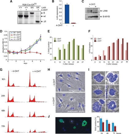
The retinoblastoma tumor suppressor protein (pRB) and related p107 and p130 "pocket proteins" function together with the E2F transcription factors to repress gene expression during the cell cycle and development. Recent biochemical studies have identified the multisubunit DREAM pocket protein complexes in Drosophila melanogaster and Caenorhabditis elegans in regulating developmental gene repression. Although a conserved DREAM complex has also been identified in mammalian cells, its physiological function in vivo has not been determined. Here we addressed this question by targeting Lin9, a conserved core subunit of DREAM. We found that LIN9 is essential for early embryonic development and for viability of adult mice. Loss of Lin9 abolishes proliferation and leads to multiple defects in mitosis and cytokinesis because of its requirement for the expression of a large set of mitotic genes, such as Plk1, Aurora A, and Kif20a. While Lin9 heterozygous mice are healthy and normal, they are more susceptible to lung tumorigenesis induced by oncogenic c-Raf than wild-type mice. Together these experiments provide the first direct genetic evidence for the role of LIN9 in development and mitotic gene regulation and they suggest that it may function as a haploinsufficient tumor suppressor.


Figures

FIG. 1.
FIG. 1.
Early embryonic lethality of Lin9 mutant mice. (A) Schematic diagram of the Lin9 wild-type locus and the trapped allele. PCR primers for genotyping are also shown. (B) The gene trap insertion leads to a C-terminally truncated LIN9 protein fused to the β-galactosidase-neomycin marker. (C) Southern blot analysis of BglII-digested genomic DNA from wild-type and heterozygous mice. (D) PCR to detect the Lin9 wild-type and knockout alleles in genomic DNA from tail biopsy specimens with primers SG662, SG885, and SG887. The primer pair SG662 and SG887 is specific for the wild-type allele. SG885 and SG887 detect the knockout allele. The position of the primers is indicated in panel A. (E) Western blot analysis showing the expression of LIN9 and the LIN9-β-geo fusion protein in Lin9+/ testes. A fusion protein of the expected molecular weight could be detected in Lin9 heterozygous (Lin9+/) mice, but it was expressed at very low levels and was only detected after long exposure of the blot. (F) Embryonic lethal phenotype of homozygous Lin9 knockout mice. Outcomes for progeny arising from intercrosses of Lin9+/ animals are shown. Footnotes indicate that genotypes were determined by Southern blotting (a), PCR (b), or TaqMan qPCR (c).
FIG. 2.
FIG. 2.
Lin9/ embryos lack the epiblast, and the ICM of Lin9/ blastocysts is not maintained in culture. (A) Histological sections of deciduae from heterozygous Lin9+/ breedings at day 7.5 dpc were stained with H&E. Also shown are results of in situ hybridization for Oct4 and H19. About one-quarter (10 out of 48) of embryos displayed abnormal morphology. Genotyping confirmed that they were of the Lin9/ genotype. (B) Deciduae from 6.5 dpc mice were stained with H&E and used for in situ hybridization of Oct4 and H19. Twelve out of 59 embryos were highly abnormal and were presumably of the Lin9/ genotype. (C) Phase-contrast microscopy of blastocysts isolated from Lin9+/ intercrosses and cultured for 7 days. Lin9/ and Lin9+/ blastocysts hatched from their zona pellucidae. The trophectoderm (TE) attached to and spread out at the bottom of wells. The ICM underwent proliferation. Homozygotes hatched and initially formed TE and ICM; however, the ICM was not maintained.
FIG. 3.
FIG. 3.
A conditional allele of Lin9. (A) Schematic diagram of the wild-type Lin9 locus and the targeted allele. Flp-mediated deletion of the selection cassette results in the floxed allele (fl), and Cre-mediated deletion of exon 7 results in the knockout (Δfl) allele. Correct targeting of Lin9 was verified by Southern blotting (data not shown). The locations of primers used for PCR genotyping are indicated. (B) Deletion of the selection cassette from the targeted Lin9 locus in vivo was achieved by crossing to a mouse strain carrying a flp transgene. Results shown are from PCR genotyping of tail DNA from crosses of Lin9+/t mice with flp transgenic mice to detect the wt, targeted (t), and the floxed (fl) alleles with primers SG893 and SG894 (top panel). Primers SG506 and SG722 were used to detect the wt and fl alleles (middle panel). The bottom panel shows a Flp-specific PCR with primers SG913 and SG914 to verify the presence of the Flp transgene. (C) Lin9fl/+ animals were crossed to mice ubiquitously expressing Cre recombinase. PCR genotyping of tail DNA with primers SG722 and SG893 was used to detect the wt, fl, and Δfl alleles. (D) Lin9Δfl results in lethality at homozygosity. Lin9Δfl/fl mice were intercrossed. Genotypes of progeny were determined by PCR.
FIG. 4.
FIG. 4.
Impaired proliferation of Lin9-deficient MEFs. (A) MEFs isolated from embryos with the indicated genotypes were treated with 4-OHT. The genotype was determined by PCR. (B and C) Reduction of Lin9 mRNA and protein levels in 4-OHT-treated MEFs as determined by RT-qPCR and immunoblotting, respectively. B-MYB protein levels are also shown and were unchanged. (D) Impaired proliferation of Lin9-deficient MEFs. Lin9+/+ CreERT2 and Lin9fl/fl CreERT2 MEFs were treated with 4-OHT where indicated, and growth was analyzed. (E) Lin9fl/fl CreERT2 MEFs were made quiescent by serum starvation. At 24 h after serum removal, 4-OHT was added, as indicated. Forty-eight hours later, reentry into the cell cycle was induced by addition of 20% serum and was monitored by BrdU incorporation. (F) Entry into mitosis of the indicated MEF cultures was analyzed by staining with an antibody specific for phosphorylated histone H3. The experiment shown in panels E and F has been repeated several times. One representative experiment is shown. (G) Lin9fl/fl CreERT2 MEFs were treated with 4-OHT for 48 h. At the indicated time points after 4-OHT treatment, the cell cycle profile was determined by flow cytometry. (H) Binucleated cells in 4-OHT-treated Lin9fl/fl Cre-ERT2 MEFs. The percentage of binucleated cells was determined by DAPI staining and fluorescence microscopy. (I) Examples of nuclear abnormalities observed upon deletion of Lin9. Cells were stained with DAPI and analyzed by fluorescence microscopy. (J) Ongoing DNA synthesis upon deletion of Lin9. Lin9fl/fl CreERT2 MEFs were treated or not with 4-OHT for 48 h. At the indicated time points thereafter, DNA synthesis was quantified by BrdU incorporation. The left panel shows abnormal morphology of BrdU-positive nuclei (green) at 48 h after 4-OHT treatment. The right panel shows the percentage of BrdU-positive cells at the indicated time points.
FIG. 5.
FIG. 5.
Mitotic phenotype and senescence of LIN9-deficient MEFs. (A) H2B-EGFP was expressed in Lin9fl/fl CreERT2 MEFs treated with or without 4-OHT. Images were recorded by time-lapse microscopy. Selected frames from the time-lapse videos are shown. The arrows point to cells whose mitosis or cytokinesis was abnormal. (B) Multipolar spindles after deletion of Lin9. Cells were stained with antibodies directed against α-tubulin (green) and γ-tubulin (red) to detect mitotic spindles and centrosomes, respectively. DNA was counterstained with DAPI (blue). (C) Senescence of Lin9-deficient MEFs. Early-passage primary Lin9fl/fl CreERT2 MEFs were treated with 4-OHT as indicated. Two weeks after 4-OHT treatment, Lin9fl/fl CreERT2 MEFs were stained for SA-β-gal, a marker of senescent cells. Phase-contrast images are shown.
FIG. 6.
FIG. 6.
Lin9 target genes. (A) Transcriptional profiling of Lin9 knockout MEFs. RNA was isolated from Lin9fl/fl CreERT2 MEFs treated with or without 4-OHT for 48 h. Genome-wide microarray analysis was used to identify LIN9-regulated genes. Expression data were stored in Array Express (http://www.ebi.ac.uk/arrayexpress/; accession number E-MEXP-2097). (B) RT-qPCR was used to confirm up- and downregulation of the indicated genes upon deletion of Lin9. (C) Genes downregulated after deletion of Lin9 with a known function in mitosis. (D) ChIP analysis of the indicated promoters with chromatin isolated from untreated and 4-OHT-treated Lin9fl/fl CreERT2 MEFs. Bound DNA was analyzed by qPCR.
FIG. 7.
FIG. 7.
Loss of Lin9 in adult mice leads to rapid loss of proliferating intestinal epithelial cells. (A) Inducible deletion of Lin9 in adult mice. (B) Genotyping of tissues from Lin9fl/fl CreERT2 mice treated with tamoxifen by intraperitonal injection for three consecutive days. Two- to 3-month-old Lin9+/+ CreERT2 or Lin9fl/fl CreERT2 mice were treated with tamoxifen. The intestinal phenotype was analyzed at the indicated day (d) after the first injection. (Upper panel) H&E staining showing normal crypt-villus morphology in the duodenum of Lin9+/+ CreERT2 animals. (Lower panels) Intestinal epithelium atrophy after deletion of Lin9 in Lin9fl/fl CreERT2 mice. (C) Intestinal epithelium atrophy is associated with the loss of proliferating cells. Mice were treated with tamoxifen as described above. At the indicated time points after the first tamoxifen injection, proliferation was analyzed by staining for the proliferation marker Ki-67. (D) Four days after the first tamoxifen injection, mice were injected with BrdU for 2 h to label proliferating cells. The percentage of BrdU-positive cells was analyzed by staining with a BrdU antibody (E) Abnormal nuclei in the intestinal epithelium upon deletion of Lin9. (Right) Morphometric analysis of nuclear size. The mean nuclear diameter of villus enterocytes increased from 23.19 μm2 to 30.14 μm2 (P < 0.0001). (F) Lin9 was deleted by three consecutive daily intraperitoneal injections of 1 mg of tamoxifen into Lin9fl/fl CreERT2 mice. Three and four days after the first injection, RNA was isolated from the intestine. Levels of the indicated mitotic genes were analyzed by real time RT-PCR.
FIG. 8.
FIG. 8.
Lin9 heterozygosity leads to a weakened spindle checkpoint and contributes to tumorigenicity in a model of non-small-cell lung cancer. (A) Lin9fl/+ CreERT2 MEFs were treated with 4-OHT or untreated. At the indicated times after treatment the percentage of binuclear cells was determined. (B) Growth of 4-OHT-treated and untreated Lin9fl/+ CreERT2 MEFs was analyzed. (C) Gene expression in Lin9fl/+ CreERT2 MEFs was analyzed by qPCR. Data show expression in 4-OHT-treated cells relative to untreated cells. (D) Mitotic index of untreated and 4-OHT-treated Lin9fl/+ CreERT2 MEFs following treatment with nocodazole for the indicated time periods. The experiment was repeated several times. One representative experiment is shown. (E) Lin9 heterozygosity results in growth in soft agar. Lin9fl/+ CreERT2 MEFs were immortalized with SV40(LT) or (LTΔ89-96). After treatment with 4-OHT to delete Lin9, cells formed colonies in soft agar. (Right) Number of colonies in 4-OHT-treated and untreated samples. (F) Reduced survival of Lin9 heterozygous mice in a model of small cell lung cancer. The Kaplan-Meier plot shows survival of Lin9+/+ BXB-Raf and Lin9+/ BXB-Raf mice. P values were calculated using the log-rank test. (G and H) Quantification of tumor area (G) and tumor incidence (H) in SP-C Lin9+/+ BXB-Raf and Lin9+/ BXB-Raf lungs from 4- to 5-month-old animals. Medians are indicated by the horizontal lines. P values were calculated using Student's t test. (I) H&E staining of lung sections from Lin9+/+ BXB-Raf and Lin9+/ BXB-Raf mice.

References

    1. Baker, D. J., K. B. Jeganathan, L. Malureanu, C. Perez-Terzic, A. Terzic, and J. M. van Deursen. 2006. Early aging-associated phenotypes in Bub3/Rae1 haploinsufficient mice. J. Cell Biol. 172:529-540. - PMC - PubMed
    1. Beall, E. L., M. Bell, D. Georlette, and M. R. Botchan. 2004. Dm-myb mutant lethality in Drosophila is dependent upon mip130: positive and negative regulation of DNA replication. Genes Dev. 18:1667-1680. - PMC - PubMed
    1. Beitel, G. J., E. J. Lambie, and H. R. Horvitz. 2000. The C. elegans gene lin-9, which acts in an Rb-related pathway, is required for gonadal sheath cell development and encodes a novel protein. Gene 254:253-263. - PubMed
    1. Bernstein, B. E., M. Kamal, K. Lindblad-Toh, S. Bekiranov, D. K. Bailey, D. J. Huebert, S. McMahon, E. K. Karlsson, E. J. Kulbokas, 3rd, T. R. Gingeras, S. L. Schreiber, and E. S. Lander. 2005. Genomic maps and comparative analysis of histone modifications in human and mouse. Cell 120:169-181. - PubMed
    1. Cotsiki, M., R. L. Lock, Y. Cheng, G. L. Williams, J. Zhao, D. Perera, R. Freire, A. Entwistle, E. A. Golemis, T. M. Roberts, P. S. Jat, and O. V. Gjoerup. 2004. Simian virus 40 large T antigen targets the spindle assembly checkpoint protein Bub1. Proc. Natl. Acad. Sci. U. S. A. 101:947-952. - PMC - PubMed

Publication types

MeSH terms