. 2010 Apr 19;189(2):223-32.
doi: 10.1083/jcb.200911018.

Regulation of exosome secretion by Rab35 and its GTPase-activating proteins TBC1D10A-C

Affiliations

Regulation of exosome secretion by Rab35 and its GTPase-activating proteins TBC1D10A-C

Chieh Hsu et al. J Cell Biol. .

Abstract

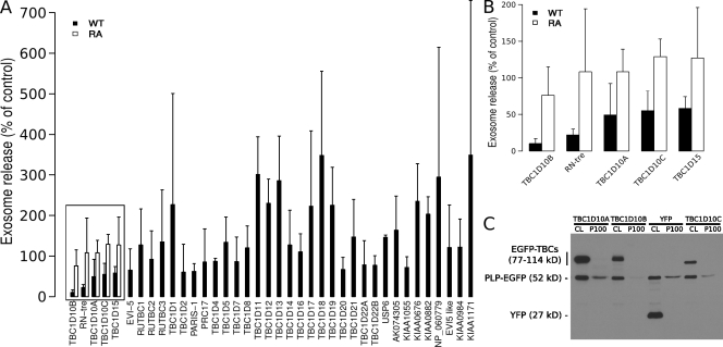
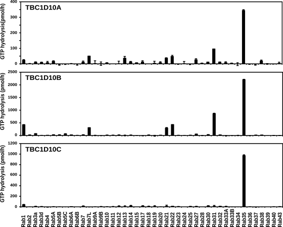
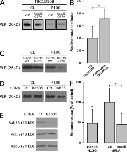
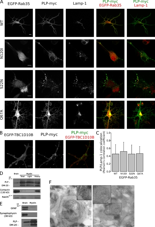
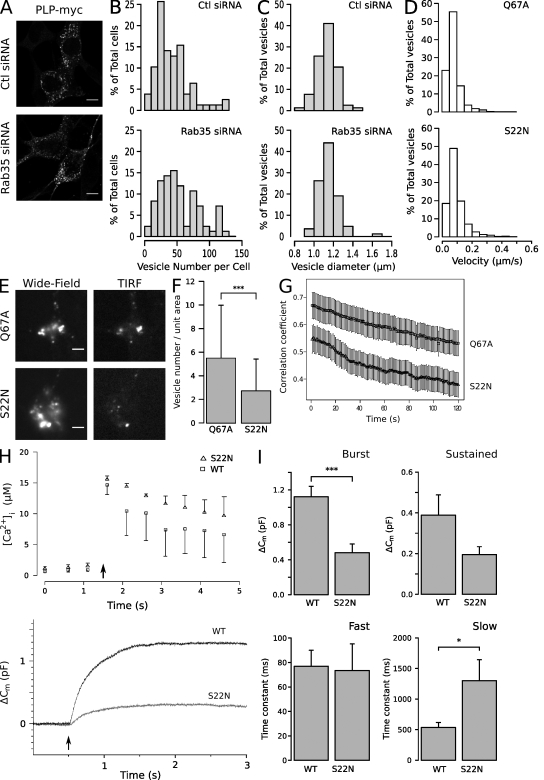
Oligodendrocytes secrete vesicles into the extracellular space, where they might play a role in neuron-glia communication. These exosomes are small vesicles with a diameter of 50-100 nm that are formed within multivesicular bodies and are released after fusion with the plasma membrane. The intracellular pathways that generate exosomes are poorly defined. Because Rab family guanosine triphosphatases (GTPases) together with their regulators are important membrane trafficking organizers, we investigated which Rab GTPase-activating proteins interfere with exosome release. We find that TBC1D10A-C regulate exosome secretion in a catalytic activity-dependent manner. We show that Rab35 is the target of TBC1D10A-C and that the inhibition of Rab35 function leads to intracellular accumulation of endosomal vesicles and impairs exosome secretion. Rab35 localizes to the surface of oligodendroglia in a GTP-dependent manner, where it increases the density of vesicles, suggesting a function in docking or tethering. These findings provide a basis for understanding the biogenesis and function of exosomes in the central nervous system.


Figures

Figure 1.
Figure 1.
Screen of a Rab GAP library identifies the TBC1D10 family as regulators of exosome secretion. (A and B) PLP-EGFP was coexpressed with a library of 38 different EGFP-tagged wild-type (WT) Rab GAPs in Oli-neu cells using a plasmid ratio of 2:1 (EGFP-TBC/PLP-EGFP). Cells transfected with both PLP-EFP and YFP were used as a reference. 16 h after transfections, the cells were switched to serum-free medium, and the medium was collected after ∼4 h of further incubation before submitting it to sequential centrifugation steps. The resulting 100,000 g pellets (exosome fraction) of each centrifugation step were analyzed by Western blotting for PLP-EGFP and the EGFP-TBCs (Rab GAPs) by anti-GFP antibodies. The mean of three independent experiments is shown in the graph. Those Rab GAPs that reduced exosome release of PLP below 60% of the control (boxed), were compared with their catalytically inactive arginine to alanine (RA) mutants, and the zoomed-in graph is shown in B. Error bars indicate the SD. (C) Western blot of the cell lysates (CL) and 100,000 g pellets (P100) is shown for one representative experiment with TBC1D10.
Figure 2.
Figure 2.
Identification of Rab35 as the target of TBC1D10A–C. Biochemical GAP assays were performed using recombinant hexahistidine-GST–tagged human Rab GTPases and hexahistidine-tagged TBC1D10 family proteins. Reactions were performed for 60 min at 37°C, using 100 pmol GST-Rab and 10 pmol hexahistidine-tagged TBC1D10A, -B, or -C. GTP hydrolysis is plotted in picomoles per hour. Error bars represent SD.
Figure 3.
Figure 3.
Inhibition of Rab35 function reduces exosome secretion of PLP. (A) Cells were transfected with PLP-myc, TBC1D10B, and either YFP or Rab35Q67A. The amounts of PLP in the cell lysates (CL) and 100,000 g pellets (P100) were quantified, and the results of two experiments performed in duplicates are shown in B (values represent the mean ± SD; *, P < 0.05; Welch’s two-sample t test). (C) Cells were transfected with PLP-myc together with either wild-type (WT) Rab35 or the dominant-negative mutant (Rab35N120I). (D and E) After delivery of control (Ctl) or siRNA against Rab35 into cells by nucleofection, the cell lysates were analyzed by Western blotting with Rab35 antibodies to monitor knockdown efficiency. Actin and Rab5 were used as controls. The amount of PLP in the cell lysates and 100,000 g pellets were determined after Rab35 RNAi. (F) Results are expressed as the mean ± SD of four experiments (*, P < 0.05; one-sample t test and Welch’s two-sample t test).
Figure 4.
Figure 4.
Localization of Rab35. (A) Oli-neu cells were transfected with PLP-myc together with EGFP-Rab35S22N, EGFP-Rab35N120I, EGFP-Rab35Q67A, or wild-type EGFP-Rab35 (WT) and analyzed by confocal microscopy. Wild-type and the GTP-locked Rab35Q67A were detected at the plasma membrane, whereas the GDP-locked Rab35S22N and the nucleotide-empty EGFP-Rab35N120I were mainly found within the cytosol and in vesicles that contained PLP and Lamp-1. (B) Oli-neu cells were cotransfected with EGFP-TBC1D10B and PLP-myc and analyzed by confocal microscopy. (C) Quantification of colocalization of PLP with Lamp-1 within 7 × 7–µm intracellular regions after coexpression with the different protein Rab35 mutants is shown (n = ∼27–38). The values represent the mean ± SD. (D) Myelin (total) and myelin subfractions (light and heavy) were purified from the brain homogenates (brain) of adult mice, and the amounts of Rab35, PLP/DM20, and contactin were determined by Western blotting. Rab35 was detected in purified myelin (total) and in the subfraction of higher density (heavy). (E) Purified myelin did not contain relevant levels of GFAP or synaptophysin proteins, confirming the purity of isolated myelin. (F) Immunoelectron microscopy analysis of myelin in the spinal cord of adult mice with PLP antibodies. The left image shows an MVB within a cytoplasmic channel of compact myelin, and the right image shows an MVB in the abaxonal space. MVBs (boxed areas) are displayed at higher magnification in the insets. Bars: (A and B) 10 µm; (F) 200 nm.
Figure 5.
Figure 5.
Rab35 functions in recruitment of the endosomal vesicle to the plasma membrane. (A) Control (Ctl) or siRNA against Rab35 was delivered into cells by nucleofection, and the cells were imaged by immunofluorescence microscopy to detect PLP-myc. (B and C) Vesicle number and size are displayed in a histogram. Note that vesicle size was unaffected, whereas the number of vesicles increased after Rab35 knockdown (n = ∼80 cells from three independent experiments). (D) To analyze vesicular movement, PLP-myc was cotransfected with wild-type or mutant EGFP-tagged Rab35, cells were stained with LysoTracker red DND-99, and time-lapse images were acquired at 1 frame every 2 s at 37°C. The motility of LysoTracker-labeled vesicles was slightly reduced when active GTP-locked Rab35Q67A was expressed (n = ∼1,800 vesicles from three independent experiments). (E and F) The mean number of LysoTracker green DND-26–labeled vesicles in a field of 12.7 µm × 12.7 µm (unit area) was determined in the evanescent excitation field. Expression of the GTP-locked Rab35Q67A increased the number of vesicles in the TIRF evanescence field as compared with the GDP-locked Rab35S22N (n = 70 cells from three independent experiments; mean ± SD; ***, P < 0.001; Welch’s two-sample t test). (G) The mobility of LysoTracker green DND-26–labeled vesicles was determined by time-lapse TIRF microscopy and analyzed by calculating the temporal correlation coefficient between pairs of images separated by time. The degree of temporal colocalization is inversely related to vesicle motility (n = ∼35 videos from three independent experiments; mean ± SEM). (H) Representative traces of intracellular Ca2+ concentration (top) and membrane capacitance (bottom) in response to flash photolysis of caged Ca2+ (indicated by arrows) in cells expressing wild-type (black trace) and GDP-locked Rab35S22N (gray trace). Error bars represent SEM. (I) A decrease in capacitance during the exocytotic burst (0–1 s) and an increase in time constant of the slow compartment were observed in cells expressing Rab35S22N (*, P < 0.05; ***, P < 0.001; Mann-Whitney test; mean ± SEM). Bars: (A) 10 µm; (E) 5 µm.

Similar articles

Cited by

References

    1. Aoki N., Jin-no S., Nakagawa Y., Asai N., Arakawa E., Tamura N., Tamura T., Matsuda T. 2007. Identification and characterization of microvesicles secreted by 3T3-L1 adipocytes: redox- and hormone-dependent induction of milk fat globule-epidermal growth factor 8-associated microvesicles. Endocrinology. 148:3850–3862 10.1210/en.2006-1479 - DOI - PubMed
    1. Barile M., Pisitkun T., Yu M.J., Chou C.L., Verbalis M.J., Shen R.F., Knepper M.A. 2005. Large scale protein identification in intracellular aquaporin-2 vesicles from renal inner medullary collecting duct. Mol. Cell. Proteomics. 4:1095–1106 10.1074/mcp.M500049-MCP200 - DOI - PMC - PubMed
    1. Barres B.A. 2008. The mystery and magic of glia: a perspective on their roles in health and disease. Neuron. 60:430–440 10.1016/j.neuron.2008.10.013 - DOI - PubMed
    1. Buschow S.I., Nolte-’t Hoen E.N., van Niel G., Pols M.S., ten Broeke T., Lauwen M., Ossendorp F., Melief C.J., Raposo G., Wubbolts R., et al. 2009. MHC II in dendritic cells is targeted to lysosomes or T cell-induced exosomes via distinct multivesicular body pathways. Traffic. 10:1528–1542 10.1111/j.1600-0854.2009.00963.x - DOI - PubMed
    1. Conde-Vancells J., Rodriguez-Suarez E., Embade N., Gil D., Matthiesen R., Valle M., Elortza F., Lu S.C., Mato J.M., Falcon-Perez J.M. 2008. Characterization and comprehensive proteome profiling of exosomes secreted by hepatocytes. J. Proteome Res. 7:5157–5166 10.1021/pr8004887 - DOI - PMC - PubMed

Publication types

MeSH terms