. 2010 Apr 19;189(2):311-23.
doi: 10.1083/jcb.200912010.

Rho1 regulates apoptosis via activation of the JNK signaling pathway at the plasma membrane

Affiliations

Rho1 regulates apoptosis via activation of the JNK signaling pathway at the plasma membrane

Amanda L Neisch et al. J Cell Biol. .

Abstract

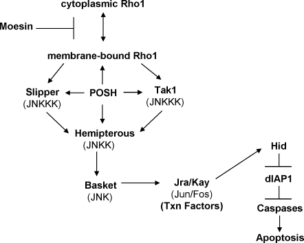
Precisely controlled growth and morphogenesis of developing epithelial tissues require coordination of multiple factors, including proliferation, adhesion, cell shape, and apoptosis. RhoA, a small GTPase, is known to control epithelial morphogenesis and integrity through its ability to regulate the cytoskeleton. In this study, we examine a less well-characterized RhoA function in cell survival. We demonstrate that the Drosophila melanogaster RhoA, Rho1, promotes apoptosis independently of Rho kinase through its effects on c-Jun NH(2)-terminal kinase (JNK) signaling. In addition, Rho1 forms a complex with Slipper (Slpr), an upstream activator of the JNK pathway. Loss of Moesin (Moe), an upstream regulator of Rho1 activity, results in increased levels of Rho1 at the plasma membrane and cortical accumulation of Slpr. Together, these results suggest that Rho1 functions at the cell cortex to regulate JNK activity and implicate Rho1 and Moe in epithelial cell survival.


Figures

Figure 1.
Figure 1.
Moe suppresses apoptosis and the canonical apoptotic cascade. (A and B) Hemizygous MoeG0323 wing discs have increased activated caspase staining (A) compared with wild-type wing discs (B). (C) The wing disc expression domain of dpp-Gal4, a driver used throughout this study, is illustrated. (D) Expression of a UAS-Moe RNAi transgene under a dpp-Gal4 driver (expression region is marked by UAS-GFP; D and D′) also induces apoptosis (merged images shown in D″). Arrowhead in D″ indicates caspase-positive, GFP-negative cells, presumably caused by the perdurance of Moe RNAi. Reducing the dosage of Rho1 (Rho172R/+) in a background expressing the UAS-Moe RNAi transgene with the dpp-Gal4 driver suppresses apoptosis (compare cells in the GFP stripe in E with D). (F) Expression of a UAS-Rho1+ transgene under the dpp-Gal4 driver also induces apoptosis. Note that in D′ and F′, the GFP-positive stripe of cells is wider than in wild type because cells that lose epithelial integrity move basally and out of the stripe of dpp-Gal4 expression. (G) In MoeG0323 wing cells fixed using TCA, cortical Rho1 protein levels are increased compared with adjacent posterior cells rescued by a UAS-Moe+ transgene driven by enGal4 (en>Moe+). The morphology of this imaginal disc is distorted because of the Moe mutation. (H) In contrast, similar expression of two copies of an activated Moe transgene (en>2X MoeT559D) results in decreased levels of cortical Rho1 protein. Arrowheads indicate the anterior–posterior compartment boundary, and posterior is to the right in all panels. (I) The wing expression domain of enGal4 is shown. For all genotypes, examples shown are representative of ≥20 imaginal discs examined. Bar, 25 µm.
Figure 2.
Figure 2.
Ectopic Rho1 expression induces JNK pathway activation and apoptosis. (A and B) Compared with wild-type discs (A), a reporter for hid, a proapoptotic gene and canonical apoptotic pathway component, is strongly up-regulated in cells that express a UAS-Rho1+ transgene (B). (C and D) puc-lacZ, a reporter for JNK signaling, is increased in dpp-Gal4; UAS-Rho1+–expressing cells. (E and F) Mmp1, an additional downstream target of JNK signaling, is also up-regulated in cells expressing the Rho1 transgene (compare E with F). (G and H) Apoptosis, caused by expression of dpp-Gal4; UAS-Rho1+ (dpp>Rho1; G) is increased by reducing the dosage of puc (H) and thereby increasing JNK signaling. (I–K) Conversely, apoptosis induced by dpp>Rho1+ is suppressed by blocking JNK signaling at the level of Bsk (UAS-bskDN [I] or UAS-puc [J]), the sole Jun kinase, or Hep (UAS-hepRNAi; K), a JNK kinase. (L) Knockdown of both Slpr and Tak1, two JNKKKs, together in dpp>Rho1+-expressing cells suppresses apoptosis to a large extent. For all genotypes, examples shown are representative of ≥20 imaginal discs examined. Bar, 25 µm.
Figure 3.
Figure 3.
Hep, Tak1, and Slpr function together with Moe and Rho1 in the adult wing. (A and B) Expression of dpp>MoeRNAi on its own slightly reduces the area in the region of expression between wing veins L3 and L4 (marked by asterisks; compare A with B) but does not otherwise affect wing morphology. (C) Reducing puc dosage alone has no phenotype. However, reducing puc dosage in the dpp>MoeRNAi background or in a dpp>Rho1 background severely reduces the wing blade (quantifications in E; example of phenotype in dpp>MoeRNAi background in D). (E) This phenotype is suppressed by reducing the level of Rho1, slpr, hep, Tak1, or POSH but not by reducing the activity of Ask1. For quantification, n > 400 wings for each genotype. (F–I) Wing phenotypes scored were rudimentary (F), serrated (G), vein defect (H), and normal (I), which has a reduction in the area between wing veins L3 and L4. Bars, 475 µm.
Figure 4.
Figure 4.
Rho1 interacts with Slpr independent of its GTP-binding state. (A) A schematic diagram of the structure of the Slpr indicating different domains and their amino acids coordinates. Lines below indicate constructs used to determine regions required for interactions with Rho1. LZ, Leu zipper domain. (B and C) S2 cells were cotransfected with expression constructs for the indicated forms of Slpr, Rho1, Cdc42, and Rac1. (B) Wild-type, CA (Rho1V14), and DN (Rho1N19) forms of Rho1 all coimmunoprecipitate with full-length Slpr protein, although Rho1N19 is consistently the strongest. (C) Deletion of the CRIB domain greatly diminishes interaction between Slpr and the CA forms of Cdc42 (Cdc42V12) and Rac1 (Rac1V12) but does not affect interaction with Rho1. (D) In vitro–translated Slpr containing the SH3, kinase, Leu zipper, and CRIB domains preferentially binds to GTP-loaded Rac1 and Cdc42-GST fusion proteins. In contrast, this domain shows no preference for GTP- versus GDP-loaded Rho1. (E–G) The kinase domain of Slpr is required for co-IP with Rho1 (E) but not for co-IP with Cdc42 (F) or Rac1 (G). (H) Endogenous Slpr from cultured S2 cells coimmunoprecipitates with DN Rho1 (Rho1N19). IB, immunoblot.
Figure 5.
Figure 5.
Rho1-induced apoptosis is dependent on its cortical localization. (A) Expression of a DN Rho1 (Rho1N19) transgene under the dpp-Gal4 driver induces apoptosis. (B) This effect requires JNK signaling, as inhibiting Bsk by expressing a DN transgene in cells expressing Rho1N19 suppresses apoptosis. (C) Expression of a Rho1 transgene with a missense mutation (C189S) in the CAAX box does not result in apoptosis. (D) Reducing the level of cortically localized Rho1 using an allele of Rho1 (Rho1E3.10) with a missense mutation (C189Y) in the CAAX box suppresses apoptosis caused by expression of the UAS-Moe RNAi transgene under dppblnk-Gal4 (dpp>MoeRNAi). Bar, 25 µm.
Figure 6.
Figure 6.
The cortical abundance of Slpr is increased in Moe mutant cells. (A) Expression of dpp>MoeRNAi results in increased apical Slpr localization. (B) A higher magnification view of the region indicated by the box in A″ is presented showing increased Slpr punctate localization in the apical domain of Moe-depleted cells. (C) Pixel intensity across the region of the disc, from anterior (A) to posterior (P), shown in B indicates that when Moe levels are knocked down, apical (1.5 µm below surface) Slpr levels are much greater than in the wild-type cells. More basally (10.5 µm below surface), Slpr levels are uniform. Bars, 25 µm.
Figure 7.
Figure 7.
A model for Moe and Rho1 function in apoptosis based on genetic, biochemical, and subcellular localization data. Moe negatively regulates Rho1 activity, at least in part by preventing its localization to the cell cortex. Cortical Rho1 forms a complex that contains Slpr, Tak1, Hep, and POSH and functions to activate the JNK pathway. Downstream of the JNK pathway, hid transcription is up-regulated to trigger apoptosis.

Similar articles

Cited by

References

    1. Adachi-Yamada T., O'Connor M.B. 2002. Morphogenetic apoptosis: a mechanism for correcting discontinuities in morphogen gradients. Dev. Biol. 251:74–90 10.1006/dbio.2002.0821 - DOI - PubMed
    1. Afshar K., Stuart B., Wasserman S.A. 2000. Functional analysis of the Drosophila diaphanous FH protein in early embryonic development. Development. 127:1887–1897 - PubMed
    1. Agnès F., Suzanne M., Noselli S. 1999. The Drosophila JNK pathway controls the morphogenesis of imaginal discs during metamorphosis. Development. 126:5453–5462 - PubMed
    1. Bangi E., Wharton K. 2006. Dpp and Gbb exhibit different effective ranges in the establishment of the BMP activity gradient critical for Drosophila wing patterning. Dev. Biol. 295:178–193 10.1016/j.ydbio.2006.03.021 - DOI - PubMed
    1. Bloor J.W., Kiehart D.P. 2002. Drosophila RhoA regulates the cytoskeleton and cell-cell adhesion in the developing epidermis. Development. 129:3173–3183 - PubMed

Publication types

MeSH terms