. 2010 Apr 19;189(2):325-38.
doi: 10.1083/jcb.200904114.

Convergence of Notch and beta-catenin signaling induces arterial fate in vascular progenitors

Affiliations

Convergence of Notch and beta-catenin signaling induces arterial fate in vascular progenitors

Kohei Yamamizu et al. J Cell Biol. .

Erratum in

  • J Cell Biol. 2013 Jul 8;202(1):179

Abstract

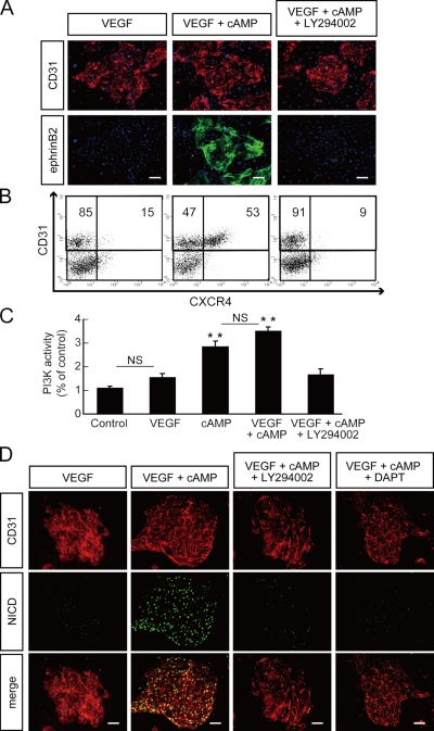
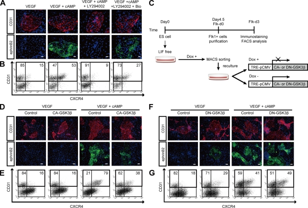
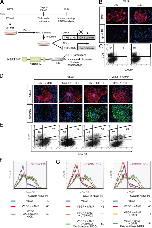
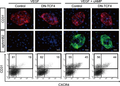
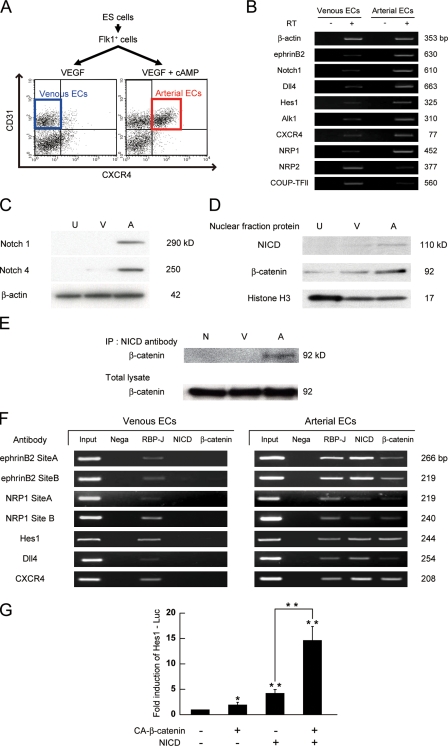
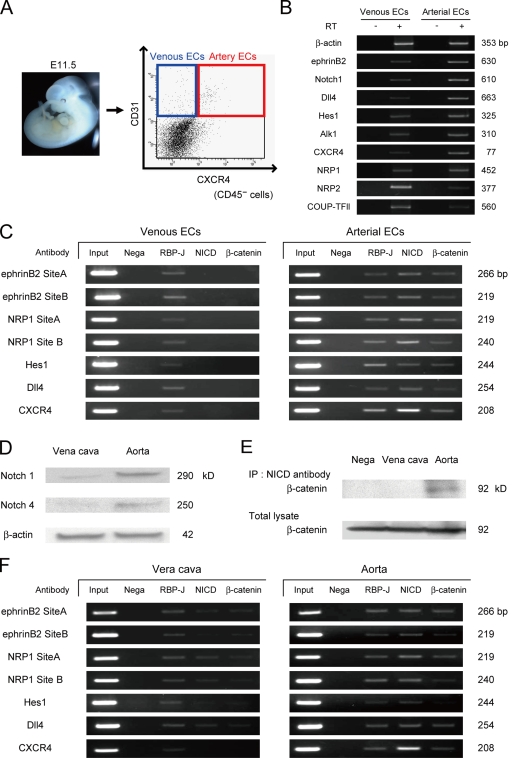
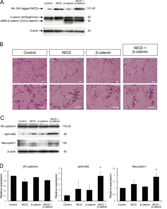
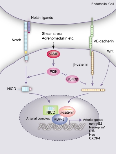
Molecular mechanisms controlling arterial-venous specification have not been fully elucidated. Previously, we established an embryonic stem cell differentiation system and demonstrated that activation of cAMP signaling together with VEGF induces arterial endothelial cells (ECs) from Flk1(+) vascular progenitor cells. Here, we show novel arterial specification machinery regulated by Notch and beta-catenin signaling. Notch and GSK3beta-mediated beta-catenin signaling were activated downstream of cAMP through phosphatidylinositol-3 kinase. Forced activation of Notch and beta-catenin with VEGF completely reconstituted cAMP-elicited arterial EC induction, and synergistically enhanced target gene promoter activity in vitro and arterial gene expression during in vivo angiogenesis. A protein complex with RBP-J, the intracellular domain of Notch, and beta-catenin was formed on RBP-J binding sites of arterial genes in arterial, but not venous ECs. This molecular machinery for arterial specification leads to an integrated and more comprehensive understanding of vascular signaling.


Figures

Figure 1.
Figure 1.
Inhibitory effect of PI3K inhibitor LY294002 on arterial EC induction from Flk1+ cells. (A) Double-fluorescent staining for CD31 and ephrinB2 after a 3-d culture of Flk1+ cells (Flk-d3). Top panels: CD31 (pan-ECs, red) and DAPI (blue). Bottom panels: EphB4-Fc (ephrinB2+ arterial ECs, green) and DAPI (blue). Flk1+ cells stimulated with VEGF alone (50 ng/ml; left panels), VEGF and 8bromo-cAMP (0.5 mM; middle panels), or VEGF, 8bromo-cAMP, and a PI3K inhibitor, LY294002 (7.5 µM; right panels). Bars: 100 µm. (B) Flow cytometry for CD31 and CXCR4 expression at Flk-d3. Percentages of CXCR4+/CD31+ arterial ECs and CXCR4/CD31+ venous ECs in total ECs (CD31+ cells) are indicated. (C) PI3K activity at Flk-d3. Flk1+ cells stimulated with vehicle, VEGF, 8bromo-cAMP, VEGF and 8bromo-cAMP, or VEGF, 8bromo-cAMP, and LY294002 (7.5 µM; n = 3; **, P < 0.01 vs. vehicle; NS: not significant). (D) Double-fluorescent immunostaining for cleaved Notch intracellular domain (NICD) and CD31 at Flk-d3. Left panels, CD31 (pan-ECs, red). Middle panels, cleaved NICD (green). Right panels, merged image. Flk1+ cells stimulated with VEGF alone, VEGF and 8bromo-cAMP, VEGF, 8bromo-cAMP and LY294002, or VEGF, 8bromo-cAMP, and a γ-secretase inhibitor, DAPT (2.5 µM). Bars: 100 µm.
Figure 2.
Figure 2.
Inhibitory effect of GSK3β on arterial EC differentiation. (A) Double-fluorescent staining for CD31 and ephrinB2 at Flk-d3. Top panels, CD31 (pan-ECs, red) and DAPI (blue). Bottom panels, EphB4-Fc (ephrinB2+ arterial ECs, green) and DAPI (blue). Flk1+ cells stimulated with VEGF alone (50 ng/ml), VEGF and 8bromo-cAMP (0.5 mM), VEGF, 8bromo-cAMP, and LY294002 (7.5 µM), or VEGF, 8bromo-cAMP, LY294002, and a GSK3β inhibitor, Bio (100 nM). Bars: 100 µm. (B) Flow cytometry for CD31 and CXCR4 expression at Flk-d3. Percentages of CXCR4+/CD31+ arterial ECs and CXCR4/CD31+ venous ECs in total ECs (CD31+ cells) are indicated. (C) Experimental system for GSK3β expression. ES cell line expressing constitutive active (CA) form or dominant-negative (DN) form of GSK3β by tetracycline-inducible expression system (Tet-Off) were established. Doxycycline (Dox) was added during the first 4.5 d of culture of ES cell differentiation to Flk1+ cells. Subsequently, Flk1+ cells were sorted by MACS and plated on type IV collagen-coated dishes, and cells were cultured in the presence or absence of 1 µg/ml Dox. (D and E) Induction of CA-GSK3β. (F and G) Induction of DN-GSK3β. (D and F) Double-fluorescent staining for CD31 and ephrinB2 at Flk-d3. Top panels, CD31 (pan-ECs, red) and DAPI (blue). Bottom panels, EphB4-Fc (ephrinB2+ arterial ECs, green) and DAPI (blue). Flk1+ cells were cultured with VEGF alone, or VEGF and 8bromo-cAMP, in the presence or absence of Dox. Bars: 200 µm. (E and G) Flow cytometry for CD31 and CXCR4 expression at Flk-d3. Percentages of CXCR4+/CD31+ arterial ECs and CXCR4/CD31+ venous ECs in total ECs (CD31+ cells) are indicated.
Figure 3.
Figure 3.
Arterial EC induction by dual activation of β-catenin and Notch signaling. (A) Experimental system for dual activation of Notch and β-catenin signaling. ES cell lines carrying CA-β-catenin regulated by Tet-Off system, and a fusion protein of N1ICD and estrogen receptor (ER), NERTΔOP, were established. CA-β-catenin was induced by depletion of Dox, and Notch activation was induced by nuclear translocation of NERTΔOP with addition of 4-hydrotamoxifen (OHT). 1 µg/ml Dox was added during the first 4.5 d of culture of ES cell differentiation to Flk1+ cells. After Flk1+ cells were sorted by MACS and plated on type IV collagen-coated dishes, cells were treated with or without Dox and/or OHT (150 ng/ml). (B and C) Activation of β-catenin together with VEGF. (B) Double-fluorescent staining for CD31 and ephrinB2 at Flk-d3. Left panels, Dox treatment. Right panels, Dox free (expression of CA-β-catenin). (C) Flow cytometry for CD31 and CXCR4 expression at Flk-d3. Percentages of CXCR4+/CD31+ arterial ECs and CXCR4/CD31+ venous ECs in total ECs (CD31+ cells) are indicated. (D–F) Dual activation of Notch and β-catenin signaling. (D) Double-fluorescent staining for CD31 and ephrinB2 at Flk-d3. Top panels, CD31 (pan-ECs, red) and DAPI (blue). Bottom panels, EphB4-Fc (ephrinB2+ arterial ECs, green) and DAPI (blue). Flk1+ cells were treated with VEGF alone (50 ng/ml), together with Dox+ (control), Dox+/OHT+ (Notch activated), or Dox/OHT+ (dual activated) condition. VEGF and 8bromo-cAMP (0.5 mM) treatment in Dox+ condition is shown as positive control. Bars: 100 µm. (E) Flow cytometry for CD31 and CXCR4 expression. Percentages of CXCR4+/CD31+ arterial ECs and CXCR4/CD31+ venous ECs in total ECs (CD31+ cells) are indicated. (F and G) Expression profile of CXCR4 in CD31+ ECs by flow cytometry. Percentages of CXCR4+ arterial ECs in total ECs are indicated. (F) VEGF treatment alone (blue line), VEGF and 8bromo-cAMP (red line), and VEGF together with dual activation of β-catenin and Notch activation (Dox, OHT+; green line) are shown. (G) VEGF treatment alone (blue line), VEGF and 8bromo-cAMP (red line), VEGF, 8bromo-cAMP, and LY294002 (7.5 µM; left panel), DAPT (2.5 µM; right panel, orange line), VEGF, 8bromo-cAMP, and LY294002 (left panel), or DAPT (right panel) together with dual activation of β-catenin and Notch activation (Dox, OHT+; green line) are shown.
Figure 4.
Figure 4.
Effects of DN-TCF4 on arterial EC induction from Flk1+ cells. (Top) Double-fluorescent staining for CD31 and ephrinB2 at Flk-d3. CD31 panels, CD31 (pan-ECs, red) and DAPI (blue). ephrinB2 panels, EphB4-Fc (ephrinB2+ arterial ECs, green) and DAPI (blue). Flk1+ cells induced from DN-TCF4 ES cell line were cultured with VEGF alone (50 ng/ml) or VEGF and 8bromo-cAMP (0.5 mM), in the presence or absence of 1 µg/ml Dox. Bars: 100 µm. (Bottom) Flow cytometry for CD31 and CXCR4 expression at Flk-d3. Percentages of CXCR4+/CD31+ arterial ECs and CXCR4/CD31+ venous ECs in total ECs (CD31+ cells) are indicated.
Figure 5.
Figure 5.
Arterial-specific formation of protein complex with RBP-J, NICD, and β-catenin. (A) Purification of arterial and venous ECs from ES cells. CXCR4+/CD31+ cells at Flk-d3 induced by VEGF (50 ng/ml) with 8bromo-cAMP (0.5 mM) and CXCR4/CD31+ cells induced by VEGF alone were purified as arterial and venous ECs, respectively. (B) RT-PCR for mRNA expression of arterial and venous EC markers in purified arterial and venous ECs induced from ES cells as indicated in panel A. (C) Western blot for protein expression of Notch1 and Notch4 in purified arterial and venous ECs. U, undifferentiated ES cells; V, venous ECs; A, arterial ECs. (D) Nuclear localization of NICD and β-catenin. A representative result of Western blot analysis for NICD and β-catenin using nuclear fraction of purified arterial and venous ECs. Anti-histone H3: nuclear fraction control. (E) Immunoprecipitation assay. Immunoblot with anti–β-catenin antibody for total cell lysates or cell lysates immunoprecipitated with anti-NICD antibody. N: negative control, immunoprecipitated with normal rabbit-IgG antibody. (F) ChIP assays for RBP-J, NICD, and β-catenin on RBP-J binding sites of arterial markers in ECs from ES cells. Input: PCR products generated using DNA from nonimmunoprecipitated chromatin as a template. Negative control: immunoprecipitated with normal rabbit-IgG antibody. RBP-J, NICD, β-catenin: immunoprecipitated chromatin with antibodies for corresponding proteins. (G) Hes1 Luciferase reporter assay. A Notch signaling reporter, Hes1-Lucifearse plasmid was transiently transfected to MACS-purified Flk1+ cells together with CA-β-catenin and/or NERTΔOP activation. After 24 h, the luciferase activities were measured (n = 3; *, P < 0.05; **, P < 0.01 vs. control or between corresponding values).
Figure 6.
Figure 6.
Formation of the arterial protein complex in the embryo and adult vessels. (A) Purification of arterial and venous ECs from the mouse embryo. Arterial ECs (CXCR4+/CD31+/CD45) and venous ECs (CXCR4/CD31+/CD45) were isolated from E11.5 embryos. (B) RT-PCR for mRNA expression of arterial and venous EC markers in purified arterial and venous ECs from E11.5 mouse embryo. (C) ChIP assays for RBP-J, NICD, and β-catenin on RBP-J binding sites of arterial markers in ECs from embryos. (D) Western blot for Notch1 and Notch4 in isolated aorta and vena cava. (E) Immunoprecipitation assay for isolated aorta and vena cava. Immunoblot with anti–β-catenin antibody for total cell lysates or cell lysates immunoprecipitated with anti-NICD antibody. (F) ChIP assays for RBP-J, NICD, and β-catenin on RBP-J binding sites of arterial markers in the aorta and vena cava.
Figure 7.
Figure 7.
Enhancement of arterial gene expression through dual activation of Notch and β-catenin during in vivo angiogenesis. Matrigel containing VEGF (100 ng/ml), heparin (10 units/ml), and adenoviral vectors (vehicle [control], HA-tagged N1ICD [NICD], and/or CA-β-catenin) were injected subcutaneously in mice. After 7 d, the mice were sacrificed and plugs were excised. (A) Western blot for HA-tagged N1ICD and CA-β-catenin in recovered cells from Matrigel plugs. (B) Hematoxylin and eosin staining of Matrigel sections. Overall appearances were not different. Invasion of blood vessels with vascular lumen and blood cells were observed. Bars: 200 µm. (C) Representative result of Western blot for VE-cadherin, ephrinB2, and Neuropilin1 in recovered cells from Matrigel plugs. (D) Quantitative evaluation of VE-cadherin (left graph), ephrinB2 (middle graph), and Neuropilin1 (right graph) protein expression in Matrigels. Relative expression normalized with β-actin expression is shown. (n = 3; *, P < 0.05 vs. control).
Figure 8.
Figure 8.
Molecular mechanisms of arterial EC specification. cAMP signaling, which could be induced by adrenomedullin, shear stress, and so on, activates Notch and β-catenin signaling through PI3K (and GSK3β) in vascular progenitors (as well as differentiating ECs). Notch and β-catenin signaling subsequently converges into a single protein complex with RBP-J, NICD, and β-catenin (arterial complex) on arterial genes. Notch signaling from Notch ligand binding and β-catenin signaling from Wnt and VE-cadherin should also participate in forming the complex. The arterial complex should play a central role in the specification of arterial cell fate in ECs.

Similar articles

Cited by

References

    1. Adams R.H., Wilkinson G.A., Weiss C., Diella F., Gale N.W., Deutsch U., Risau W., Klein R. 1999. Roles of ephrinB ligands and EphB receptors in cardiovascular development: demarcation of arterial/venous domains, vascular morphogenesis, and sprouting angiogenesis. Genes Dev. 13:295–306 10.1101/gad.13.3.295 - DOI - PMC - PubMed
    1. Ara T., Tokoyoda K., Okamoto R., Koni P.A., Nagasawa T. 2005. The role of CXCL12 in the organ-specific process of artery formation. Blood. 105:3155–3161 10.1182/blood-2004-07-2563 - DOI - PubMed
    1. Artavanis-Tsakonas S., Rand M.D., Lake R.J. 1999. Notch signaling: cell fate control and signal integration in development. Science. 284:770–776 10.1126/science.284.5415.770 - DOI - PubMed
    1. Cattelino A., Liebner S., Gallini R., Zanetti A., Balconi G., Corsi A., Bianco P., Wolburg H., Moore R., Oreda B., et al. 2003. The conditional inactivation of the beta-catenin gene in endothelial cells causes a defective vascular pattern and increased vascular fragility. J. Cell Biol. 162:1111–1122 10.1083/jcb.200212157 - DOI - PMC - PubMed
    1. Christensen S., Kodoyianni V., Bosenberg M., Friedman L., Kimble J. 1996. lag-1, a gene required for lin-12 and glp-1 signaling in Caenorhabditis elegans, is homologous to human CBF1 and Drosophila Su(H). Development. 122:1373–1383 - PubMed

Publication types

MeSH terms

Substances