. 2010 May 4;107(18):8363-8.
doi: 10.1073/pnas.0911378107. Epub 2010 Apr 19.

Inhibition of Mac-1 (CD11b/CD18) enhances tumor response to radiation by reducing myeloid cell recruitment

Affiliations

Inhibition of Mac-1 (CD11b/CD18) enhances tumor response to radiation by reducing myeloid cell recruitment

G-One Ahn et al. Proc Natl Acad Sci U S A. .

Abstract

Despite recent advances in radiotherapy, loco-regional failures are still the leading cause of death in many cancer patients. We have previously reported that bone marrow-derived CD11b(+) myeloid cells are recruited to tumors grown in irradiated tissues, thereby restoring the vasculature and tumor growth. In this study, we examined whether neutralizing CD11b monoclonal antibodies could inhibit the recruitment of myeloid cells into irradiated tumors and inhibit their regrowth. We observed a significant enhancement of antitumor response to radiation in squamous cell carcinoma xenografts in mice when CD11b antibodies are administered systemically. Histological examination of tumors revealed that CD11b antibodies reduced infiltration of myeloid cells expressing S100A8 and matrix metalloproteinase-9. CD11b antibodies further inhibited bone marrow-derived cell adhesion and transmigration to C166 endothelial cell monolayers and chemotactic stimuli, respectively, to levels comparable to those from CD11b knockout or CD18 hypomorphic mice. Given the clinical availability of humanized CD18 antibodies, we tested two murine tumor models in CD18 hypomorphic or CD11b knockout mice and found that tumors were more sensitive to irradiation when grown in CD18 hypomorphic mice but not in CD11b knockout mice. When CD18 hypomorphism was partially rescued by reconstitution with the wild-type bone marrow, the resistance of the tumors to irradiation was restored. Our study thus supports the rationale of using clinically available Mac-1 (CD11b/CD18) antibodies as an adjuvant therapy to radiotherapy.


Conflict of interest statement

The authors declare no conflict of interest.

Figures
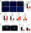
Fig. 1.
Fig. 1.
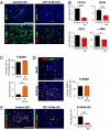
Local irradiation inhibits angiogenesis. (A) Staining of FaDu tumors grown in immunodeficient mice that were not irradiated (control, 0 Gy), irradiated with 20 Gy and harvested at day 14 postirradiation [IR 20 Gy (d14)], or recurrent after 20 Gy of irradiation [recurrent (2 mo)]. The tumors were stained for CD31 (red) and α-SMA (green) (Upper), and for CD11b (green) (Lower). Nuclei were stained with DAPI and are shown in blue. (Scale bars, 100 μm.) The bar graph shows the mean tumor volume. (B) Quantification of CD31, α-SMA, CD11b, and CD45 shown in A. Symbols and error bars in A and B are the mean ± SEM for n ≥ 5 animals per group. *, **, and *** denote P < 0.05, < 0.01, and < 0.001 by one-way ANOVA, respectively. (C) Immunostaining of matrigel implanted in mice as in A, stained with CD31 (red) and α-SMA (green) antibodies. Hoechst 33342, a diffusion dye injected immediately before the matrigel harvest, is shown in blue. (Scale bar, 100 μm.) (D) Quantification of the matrigel section from C as in B. Symbols and error bars are the mean ± SEM for n = 4 animals per group. ***, P < 0.001 by Student's t test.
Fig. 2.
Fig. 2.
CD11b monoclonal antibody treatment enhances tumor response to radiation. (A) Growth of irradiated FaDu tumors with 12 Gy (Left) or 20 Gy (Right) in mice treated with isotype control antibodies (control), Gr-1 antibodies (Gr-1 Ab), or CD11b antibodies (CD11b Ab) at 100 μg per mouse from the fourth day following irradiation for every 2 days. (B) Photographs of mice bearing FaDu tumors that had been irradiated with 20 Gy and treated with isotype control antibodies (control, Left) or CD11b antibodies (CD11b Ab, Right) for up to 2 months. Tumors had regrown in the control group (black arrowheads), whereas they became not palpable in the CD11b Ab group (black arrowheads indicate where the tumor had been originally implanted). (C) Growth of irradiated SCCVII tumors in C3H/HeJ mice with 15 Gy, followed by the control or CD11b antibodies. Symbols and error bars in A and C are the mean ± SEM for n ≥ 7 per group.
Fig. 3.
Fig. 3.
CD11b monoclonal antibodies inhibit tumor infiltrating myeloid cells expressing S100A8 and MMP-9. (A) Immunostaining of FaDu tumors from Fig. 2A (20 Gy) harvested at 7 days (d7) after irradiation, stained for CD11b+ cells using CD11b (control tumors) or anti-rat (CD11b Ab-treated tumors) antibodies (Upper). (Lower) CD31 (red) and α-SMA (green) staining. Nuclei are shown in blue with DAPI staining. (B) Quantification of immunostaining in A as area densities for CD11b, CD45, CD31, and α-SMA. (C) Quantification of S100A8 immunostaining (Upper) and colocalization with CD11b (Lower) in unirradiated (No IR) or recurrent (IR 20 Gy) tumors in Fig. 1A. (D) Immunostaining of S100A8 (red) and MMP-9 (green) in unirradiated or recurrent tumors as shown in C. Quantification of colocalization between S100A8+ cells and MMP-9 is shown in the bar graph. (E) Immunostaining of d 7 FaDu tumors as shown in A for S100A8 (red) and CD11b (green; for control tumors), or anti-rat (green; for CD11b Ab-treated tumors) antibodies. The bar graph shows quantification of S100A8 immunostaining. (Scale bars for A, D, and E: 100 μm.) Symbols and error bars in B to E are the mean ± SEM for n ≥ 3 mice per group. *, **, and *** denote P < 0.05, < 0.01, and < 0.001 by Student's t test, respectively.
Fig. 4.
Fig. 4.
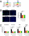
CD11b antibodies inhibit adhesion and transmigration of bone marrow-derived cells. (A) Irradiated C166 endothelial cells analyzed by FACS showed up-regulation of ICAM-1 expression in a dose- (0–20 Gy) and time- (24 h, Left; 48 h, Right) dependent manner. (B) Fluorescent images of adhered CFSE-labeled bone marrow cells (green) onto C166 endothelial cell monolayers (nuclei of the endothelial cells are shown in blue by DAPI staining) that were either pretreated with isotype control (control) or CD11b antibodies (CD11b Ab; Upper), or isolated from CD11b KO mice or CD18 hypomorphic (CD18 hypo) mice (Lower). (Scale bar, 100 μm.) Quantification of the number of CFSE-positive cells per field is shown on the right. (C) CFSE-labeled bone marrow derived cells as shown in B were incubated in modified Boyden chambers containing the culture media supplemented with no chemokine (no treatment), 10% serum, VEGF, or M-CSF. Symbols and error bars in B and C are the mean ± SEM for triplicate determinations in three independent experiments. ***, P < 0.001 by one-way ANOVA.
Fig. 5.
Fig. 5.
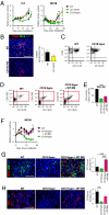
CD18 hypomorphism influences tumor response to radiation. (A) Growth of irradiated LLC (Left) or MC38 (Right) tumors with 15 Gy in the WT, CD11b KO (CD11b KO), or CD18 hypomorphic (CD18 hypo) mice. (B) Immunostaining of irradiated LLC in WT or CD11b KO as in A for S100A8 (red) and CD11b (green). DAPI shows nuclear staining in blue. Quantification of S100A8 area densities is shown in the bar graph. (C) FACS plots showing CFSE-labeled bone marrow cells isolated from WT or CD18 hypo mice for CD11b surface expression. (D) FACS analyses of the peripheral blood obtained from WT, CD18 hypo, or CD18 hypomorphic mice reconstituted with the WT bone marrow cells (CD18 hypo + WT). (E) Quantification of propidium iodide-negative, live CD18+ cells (as highlighted in the red boxes in D) in the WT, CD18 hypo, or CD18 hypo + WT mice. (F) MC38 tumor growth after irradiation with 15 Gy in WT, CD18 hypo, or CD18 hypo + WT mice shown in D and E. (G and H) Immunostaining of MC38 tumors in F for CD11b (red, G), CD18 (red, H), and CD45 (green, G). Nuclei are shown in blue with DAPI staining. (Scale bars in B, G, and H: 100 μm.) The symbols and error bars represent the mean ± SEM for n ≥ 5 per group (for A, E, and F) or n ≥ 3 per group (for B, G, and H). ** and *** denote for P < 0.01 and < 0.001, respectively, determined by one-way ANOVA.

References

    1. Verellen D, et al. Innovations in image-guided radiotherapy. Nat Rev Cancer. 2007;7:949–960. - PubMed
    1. Cummings B, et al. Five year results of a randomized trial comparing hyperfractionated to conventional radiotherapy over four weeks in locally advanced head and neck cancer. Radiother Oncol. 2007;85:7–16. - PubMed
    1. Ahn GO, Brown JM. Matrix metalloproteinase-9 is required for tumor vasculogenesis but not for angiogenesis: Role of bone marrow-derived myelomonocytic cells. Cancer Cell. 2008;13:193–205. - PMC - PubMed
    1. Lewis JS, Landers RJ, Underwood JC, Harris AL, Lewis CE. Expression of vascular endothelial growth factor by macrophages is up-regulated in poorly vascularized areas of breast carcinomas. J Pathol. 2000;192:150–158. - PubMed
    1. Shojaei F, et al. Bv8 regulates myeloid-cell-dependent tumour angiogenesis. Nature. 2007;450:825–831. - PubMed

Publication types

MeSH terms

LinkOut - more resources