. 2010 May 4;107(18):8463-8.
doi: 10.1073/pnas.1000838107. Epub 2010 Apr 19.

Domain organization and function in GluK2 subtype kainate receptors

Affiliations

Domain organization and function in GluK2 subtype kainate receptors

Utpal Das et al. Proc Natl Acad Sci U S A. .

Abstract

Glutamate receptor ion channels (iGluRs) are excitatory neurotransmitter receptors with a unique molecular architecture in which the extracellular domains assemble as a dimer of dimers. The structure of individual dimer assemblies has been established previously for both the isolated ligand-binding domain (LBD) and more recently for the larger amino terminal domain (ATD). How these dimers pack to form tetrameric assemblies in intact iGluRs has remained controversial. Using recently solved crystal structures for the GluK2 kainate receptor ATD as a guide, we performed cysteine mutant cross-linking experiments in full-length tetrameric GluK2 to establish how the ATD packs in a dimer of dimers assembly. A similar approach, using a full-length AMPA receptor GluA2 crystal structure as a guide, was used to design cysteine mutant cross-links for the GluK2 LBD dimer of dimers assembly. The formation of cross-linked tetramers in full-length GluK2 by combinations of ATD and LBD mutants which individually produce only cross-linked dimers suggests that subunits in the ATD and LBD layers swap dimer partners. Functional studies reveal that cross-linking either the ATD or the LBD inhibits activation of GluK2 and that, in the LBD, cross-links within and between dimers have different effects. These results establish that kainate and AMPA receptors have a conserved extracellular architecture and provide insight into the role of individual dimer assemblies in activation of ion channel gating.


Conflict of interest statement

The authors declare no conflict of interest.

Figures
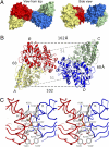
Fig. 1.
Fig. 1.
Crystal structure of the GluK2 ATD tetramer. (A) Molecular surface viewed from the top (Left) and after rotation by 90° (Right) with the four subunits colored individually. (B) Ribbon diagram showing how the GluK2 ATD AB and CD subunit dimers pack via the lateral edges of domain R2 to form a tetramer; the CA atoms of Thr2 and Glu384, which define the N and C termini of the ATD in each subunit, are indicated by colored spheres connected by dashed lines; black dumbbells indicate the CA positions of L151 in the AB and CD dimers. (C) Stereo view of the interaction surface connecting subunits B and D in the GluK2 ATD tetramer. Side chains that mediate intersubunit contacts are represented as sticks; orange spheres indicate the CA positions of Gly215. The view matches that in the right panel in A.
Fig. 2.
Fig. 2.
GluK2 ATD cross-links created by cysteine mutagenesis. (A) The blots show (left to right) spontaneous oligomerization for wild-type GluK2; the C540Y/C545V/C564S 3× (–) triple mutant; and the 4× (–) mutant which contained in addition the exchange C827N. The additional change, C840Q, to produce the 5× (–) mutant yielded a construct which ran as a monomer with the same mobility as wild-type GluK2 under reducing conditions. (B) The L151C mutation in the AB subunit ATD dimer interface produced spontaneous dimer formation (control, Con), which was not enhanced by incubation with CuPhen (oxidizing condition, Ox) and which was reversed by incubation with 10 mM BME (reducing condition, Red). (C) The G215C mutant in the BD subunit interdimer interface also produced spontaneous dimer formation which was not enhanced by incubation with CuPhen. (D) The M214C/Y240C double mutant, designed to cross-link helices I and K in the BD subunit tetramer interface, also produced dimer formation. (E) The L151C/ G215C double mutant showed bands for both dimers and tetramers. (F) Outside-out patch responses for GluK2 5× (–) had kinetics similar to wild-type GluK2; in oxidizing conditions the current in CuPhen (red curve; monoexponential fit, kdes = 131 s−1) was 88% of the peak current in DTT (blue curve; kdes = 117 s−1), but on average, peak currents in CuPhen were 101 ± 14% of the amplitude of those in DTT. (G) In reducing conditions; 10 mM glutamate activated rapidly desensitizing currents for the L151C mutant (blue fitted curve; kdes = 100 s−1). The peak amplitude was inhibited in oxidizing conditions (red fitted curve; kdes = 102 s−1; current in 10 μM CuPhen was 49 ± 7% of that in 10 mM DTT, P = 3%, n = 6 patches). The inhibition was readily reversible (green fitted curve; kdes = 89 s−1). (H) The G215C mutant also was reversibly inhibited in oxidizing conditions (red fitted curve; kdes = 63 s−1); control and wash responses in DTT were similar (blue and green curves; kdes = 61 s−1 and kdes = 60 s−1, respectively). The inhibition was statistically significant; peak current in CuPhen was 55 ± 8% of the DTT current (P < 0.05, n = 6). (I) L151C-G215C double mutants were more strongly inhibited in oxidizing conditions (red fitted curve; kdes = 74 s−1; peak amplitude in CuPhen was 35 ± 4% of that in DTT, n = 4). The inhibition was fully reversible (green fitted curve; kdes = 74 s−1).
Fig. 3.
Fig. 3.
GluK2 LBD cross-links created by cysteine mutagenesis. (A) Molecular surface of the GluA2 LBD tetramer (11) with each subunit colored using the same scheme as for the GluK2 ATD tetramer shown in Fig. 1. (B) Ribbon diagram for a homology model of the GluK2 LBD tetramer showing cysteine mutants designed to cross the A and C subunits and the A and B subunits in a tetramer assembly. (C) Western blot analysis for cross-links connecting the GluK2 LBD tetramer interface. The K667C mutation, which cross-links the loop connecting helices F and G in the AC subunit interdimer interface, produced spontaneous dimer formation, which was slightly enhanced by incubation with CuPhen (Ox) and reversed by 10 mM BME (Red). The S669C mutation at the N terminus of helix G also produced cross-linking of subunits in the AC subunit interdimer interface. The A676C/G770C double mutant, which cross-links subunits A and B in the LBD tetramer, produced weak spontaneous dimer formation, which was strongly enhanced by incubation with CuPhen. A similar effect was observed for the S680C/N771C double mutant, which also cross-links subunits A and B. (D) The double mutant L151C/K667C, which cross-links both the AB and CD subunit ATD dimer interfaces and the AC subunit dimer interface in the LBD, produced spontaneous dimer and tetramer formation, which was reversed by 10 mM BME; the cartoon shows the intersubunit connections in this mutant.
Fig. 4.
Fig. 4.
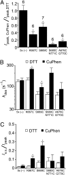
Effects of LBD cross-links on GluK2 receptor function. (A) The K667C mutant showed partial inhibition by oxidation. In this patch, the peak current amplitude was reduced from 153 pA to 30 pA, and the rate of desensitization was similar in DTT and CuPhen (kdes = 188 s−1 and 152 s−1, blue and red curves, respectively); the extent of desensitization was reduced in CuPhen, so that relative to the peak steady-state currents were 5.3 ± 0.7-fold larger than in DTT. K667C responses frequently showed rundown, as illustrated by incomplete recovery of the peak amplitude upon return to DTT (green curve; kdes = 187 s−1). (B) The S669C mutant exhibited fast desensitization in reducing conditions (blue curve; kdes 72 s−1) that was slightly, but significantly, accelerated in oxidizing conditions (red curve; kdes 100 s−1). As for the K667C mutant, the response was only partially (39% in this example) inhibited by oxidation. Desensitization was blocked substantially, with the steady-state current increasing to 19% of the peak in oxidizing conditions, compared with 2% in 10 mM DTT. (C) Glutamate-activated currents for the S680C-N771C mutant were much more strongly inhibited in CuPhen (peak amplitude 9% in this example); the inhibition was rapidly and readily reversible, as shown by blue- and green-colored exponential fits for control and recovery responses, respectively.
Fig. 5.
Fig. 5.
Bar plots illustrating kinetic effects of cross-links in the LBD. (A) Oxidizing conditions producing cross-links between AC and AB subunits give rise to distinct patterns of current inhibition. The 5× (–) construct was not inhibited by oxidation. Intermediate inhibition was observed for the K667C and S669C mutants, and much stronger reduction of the current was seen for the S680C-N771C and the A676C-G770C double mutants; number of patches indicated above bars. (B) Oxidation produced small increases in desensitization rate for all mutants, but the effect was statistically significant only for the K667C and S669C mutants (P = 0.03 for each, randomization test). Only a subset of responses in CuPhen for S680C-N771C (three of six patches) and A676C-G770C (four of six patches) were large enough to measure the desensitization rate accurately. (C) The steady-state current was strongly and significantly increased in amplitude for K667C and S669C but not for the AB subunit cross-linking mutants. *, P, < 0.05.

Similar articles

Cited by

References

    1. Dingledine R, Borges K, Bowie D, Traynelis SF. The glutamate receptor ion channels. Pharmacol Rev. 1999;51:7–61. - PubMed
    1. Unwin N. Refined structure of the nicotinic acetylcholine receptor at 4A resolution. J Mol Biol. 2005;346:967–989. - PubMed
    1. Hilf RJ, Dutzler R. X-ray structure of a prokaryotic pentameric ligand-gated ion channel. Nature. 2008;452:375–379. - PubMed
    1. Bocquet N, et al. X-ray structure of a pentameric ligand-gated ion channel in an apparently open conformation. Nature. 2009;457:111–114. - PubMed
    1. Jasti J, Furukawa H, Gonzales EB, Gouaux E. Structure of acid-sensing ion channel 1 at 1.9 A resolution and low pH. Nature. 2007;449:316–323. - PubMed

Publication types

LinkOut - more resources