. 2010 May 4;107(18):8428-33.
doi: 10.1073/pnas.1000640107. Epub 2010 Apr 19.

Palmitoylation stabilizes unliganded rod opsin

Affiliations

Palmitoylation stabilizes unliganded rod opsin

Akiko Maeda et al. Proc Natl Acad Sci U S A. .

Abstract

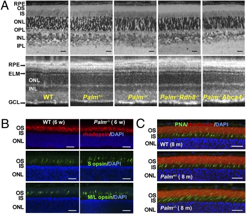
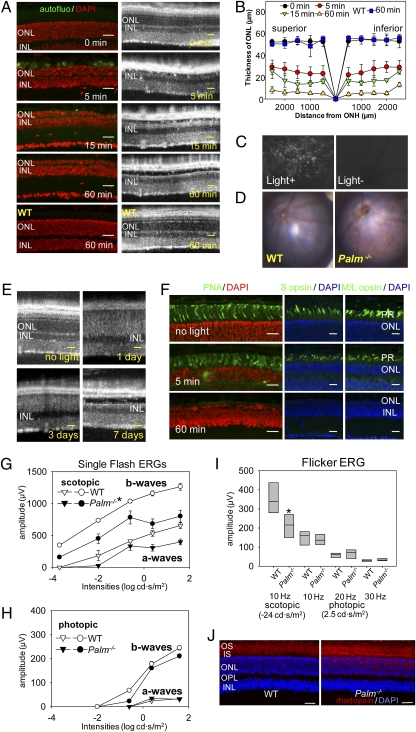
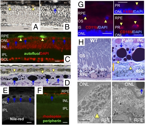
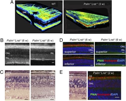
S-palmitoylation is a conserved feature in many G protein-coupled receptors (GPCRs) involved in a broad array of signaling processes. The prototypical GPCR, rhodopsin, is S-palmitoylated on two adjacent C-terminal Cys residues at its cytoplasmic surface. Surprisingly, absence of palmitoylation has only a modest effect on in vitro or in vivo signaling. Here, we report that palmitoylation-deficient (Palm(-/-)) mice carrying two Cys to Thr and Ser mutations in the opsin gene displayed profound light-induced retinal degeneration that first involved rod and then cone cells. After brief bright light exposure, their retinas exhibited two types of deposits containing nucleic acid and invasive phagocytic macrophages. When Palm(-/-) mice were crossed with Lrat(-/-) mice lacking lecithin:retinol acyl transferase to eliminate retinoid binding to opsin and thereby rendering the eye insensitive to light, rapid retinal degeneration occurred even in 3- to 4-week-old animals. This rapid degeneration suggests that nonpalmitoylated rod opsin is unstable. Treatment of 2-week-old Palm(-/-)Lrat(-/-) mice with an artificial chromophore precursor prevented this retinopathy. In contrast, elimination of signaling to G protein in Palm(-/-)Gnat1(-/-) mice had no effect, indicating that instability of unpalmitoylated opsin lacking chromophore rather than aberrant signal transduction resulted in retinal pathology. Together, these observations provide evidence for a structural role of rhodopsin S-palmitoylation that may apply to other GPCRs as well.


Conflict of interest statement

The authors declare no conflict of interest.

Figures

Fig. 1.
Fig. 1.
Palmitoylation does not affect retinal morphology of mice with different genetic backgrounds maintained under normal laboratory lighting conditions. (A) Representative retinal histology (Upper) and B-scanned averaged SD-OCT images (Lower) are shown for WT, Palm+/−, Palm−/−, Palm−/−Rdh8−/−, and Palm−/−Abca4−/− mice at 6 weeks of age (n > 3). GCL, ganglion cell layer; IPL, inner plexiform layer; INL, inner nuclear layer; OPL, outer plexiform layer; ELM, external limiting membrane; IS, inner segment; OS, outer segment; RPE, retinal pigmented epithelium. (Scale bars: 10 μm.) (B) Immunohistochemistry of 6-week-old WT and Palm−/− retinas stained with antirhodopsin (1D4), anti–S opsin and anti–M/L opsin antibodies. No degeneration was observed. Abbreviations are the same as in A. (Scale bars: 10 μm). (C) Immunohistochemistry of 8-month-old WT, Palm+/−, and Palm−/− retinas stained with peanut agglutinin lectin (PNA) and antirhodopsin (1D4) antibody. No degeneration was observed. (Scale bars: 10 μm.)
Fig. 2.
Fig. 2.
Bright light induces severe retinal degeneration in Palm−/− mice. (A) Palmitoylation-deficient (Palm−/−) and WT mice at 6 weeks of age were exposed to 10,000 lx light for periods indicated and then kept in the dark for 7 d, at which time SD-OCT and histological examinations were performed. Severe retinal degeneration was observed in Palm−/− mice whereas no degeneration was detected in WT retinas exposed to bright light for 60 min. INL, inner nuclear layer. (Scale bars: 20 μm.) (B) The thickness of the outer nuclear layer was measured 7 d after illumination with 10,000 lx for the indicated periods. ONH, optic nerve head. Error bars indicate SD of the means (n > 3). (C) Shown are representative outer retinal images obtained by SLO from 6-week-old Palm−/− mice exposed to 10,000 lx light for 5 min and then kept in the dark until examined 7 d later (Left) or from unexposed control mice kept in the dark (Right). Numerous autofluorescent retinal deposits are observed in illuminated Palm−/− mice. (D) Representative fundus images are shown from 6-week-old WT and Palm−/− mice taken 7 d after retinal illumination with 10,000 lx light for 60 min. Palm−/− mice manifest widespread atrophic changes. (E) SD-OCT B-scan imaging of the same eye performed at 1, 3, and 7 d after retinal exposure of 6-week-old Palm−/− mice to 10,000 lx light for 30 min. Hazy changes of photoreceptor layers 1 and 3 d after illumination indicate ongoing photoreceptor cell death. (Scale bars: 10 μm.) (F) Immunohistochemistry of cone photoreceptors performed with PNA, anti–S cone opsin, and anti–M/L cone opsin antibodies 7 d after retinal illumination of 6-week-old Palm−/− mice with 10,000 lx light for 5 and 60 min. (Scale bars: 10 μm.) Palm−/− and WT mice at 3 months of age were kept under 12-h light (1,000 lx)/12-h dark conditions for 4 weeks, and then ERG and histological examinations were done 7 d after dark adaptation. Full-field ERG responses were recorded under scotopic (G) and photopic (H) conditions. Both a- and b-wave amplitudes under scotopic conditions were attenuated in Palm−/− mice compared with WT animals. (I) Flicker ERGs recorded at 10, 20, and 30 Hz showed significant decreases for Palm−/− compared with WT mice under scotopic conditions, whereas no differences were observed under photopic conditions. Error bars indicate SE of the means (n > 3; *P < 0.03) vs. WT animals. (J) Retinal structures were assessed with outer segment (red, antirhodopsin 1D4), and nuclear (blue, DAPI) staining. Whereas retinal structure was preserved in both WT and Palm−/− mice, mislocalization of some rhodopsin in the ONL was observed in Palm−/− mice. Representative images are presented (n > 3). (Scale bars: 10 μm.)
Fig. 3.
Fig. 3.
Retinal deposits and macrophage infiltration in Palm−/− mice. Six-week-old Palm−/− mice with pupils dilated with 1% tropicamide were illuminated with 10,000 lx light for 60 min and then dark-adapted for 7 d before analyses. Bright field cryosection images revealed yellowish (yellow arrowheads) (A) and light blue (blue arrow) (B) retinal deposits between the ONL and the RPE. INL, inner nuclear layer; IPL, inner plexiform layer; GCL, ganglion cell layer. (Scale bars: 10 μm.) (C) Yellowish retinal deposits in A were autofluorescent (green) and blue deposits in B stained with DAPI (red). (Scale bar: 10 μm.) (D) Retinal deposits with yellow color in A were located in invasive cells at the interface of the RPE and the ONL in epon-embedded sections. Toluidine blue staining shows dark purple coloring of the blue deposits in B. (Scale bar: 10 μm.) (E) Retinal deposits with blue color in B were not stained by Nile red dye, which recognizes lipids. (Scale bars: 10 μm.) (F) The retinal deposit with blue color in B was not recognized by antirhodopsin or antiperipherin antibodies. (Scale bar: 10 μm.) (G) Yellow deposits in A were found in cells located between the outer segment (OS) and RPE that stained with macrophage markers, CD11b (Left), F4/80 (Upper Right), and CD169 (Lower Right). PR, photoreceptors. (Scale bar: 10 μm.) (H) Higher magnified images of a photoreceptor/RPE junction in WT and Palm−/− retina are shown (Upper). Yellow arrowheads and blue arrows indicate yellow and blue deposits in A and B. EM images of the area indicated as an orange bar in Palm−/− retina (Upper Right) indicate that infiltrated macrophages contained photoreceptor debris (Lower Left), and there were no photoreceptors between the RPE and the neural retina (Lower Right). OS, outer segment; INL, inner nuclear layer. (Scale bars: 3 μm.)
Fig. 4.
Fig. 4.
Absence of retinal ligand causes rapid rod photoreceptor degeneration in young Palm−/−Lrat−/− mice. (A) Three-dimensional OCT images of WT and Palm−/−Lrat−/− 3-week-old mouse retinas show reduced thickness of Palm−/− ONL (Right). (B) B-scan SD-OCT images reveal reduced thickness of ONL in Palm−/−Lrat−/− retinas, whereas Palm+/−Lrat−/− retinas exhibit a healthy structure in 6-week-old mice. (Scale bars: 10 μm.) (C) Retinal histology displays a nearly normal Palm+/−Lrat−/− retina but obvious photoreceptor degeneration in a Palm−/−Lrat−/− retina from 6-week-old mice. (Scale bars: 10 μm.) (D) Immunohistochemistry with PNA and antirhodopsin antibody reveals lack of rhodopsin expression in Palm−/−Lrat−/− retinas from 6-week-old mice. Fewer cone photoreceptors are observed in both Palm+/−Lrat−/− and Palm−/−Lrat−/− retinas, especially in the inferior retina. (Scale bars: 10 μm.) (E) Morphology and immunohistochemistry with PNA and antirhodopsin antibody staining of developing retinas from 2-week-old Palm−/−Lrat−/− mice are presented. Rhodopsin is clearly visible in the OS. (Scale bars: 10 μm.)

References

    1. Mirzadegan T, Benkö G, Filipek S, Palczewski K. Sequence analyses of G-protein-coupled receptors: similarities to rhodopsin. Biochemistry. 2003;42:2759–2767. - PMC - PubMed
    1. Mustafi D, Palczewski K. Topology of class A G protein-coupled receptors: insights gained from crystal structures of rhodopsins, adrenergic and adenosine receptors. Mol Pharmacol. 2009;75:1–12. - PMC - PubMed
    1. Torrecilla I, Tobin AB. Co-ordinated covalent modification of G-protein coupled receptors. Curr Pharm Des. 2006;12:1797–1808. - PubMed
    1. Palczewski K. G protein-coupled receptor rhodopsin. Annu Rev Biochem. 2006;75:743–767. - PMC - PubMed
    1. Ablonczy Z, Kono M, Knapp DR, Crouch RK. Palmitylation of cone opsins. Vision Res. 2006;46:4493–4501. - PMC - PubMed

Publication types

LinkOut - more resources