

Targeting stents with local delivery of paclitaxel-loaded magnetic nanoparticles using uniform fields
- PMID: 20404175
- PMCID: PMC2889533
- DOI: 10.1073/pnas.0909506107
Targeting stents with local delivery of paclitaxel-loaded magnetic nanoparticles using uniform fields
Abstract
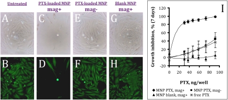
The use of stents for vascular disease has resulted in a paradigm shift with significant improvement in therapeutic outcomes. Polymer-coated drug-eluting stents (DES) have also significantly reduced the incidence of reobstruction post stenting, a disorder termed in-stent restenosis. However, the current DESs lack the capacity for adjustment of the drug dose and release kinetics to the disease status of the treated vessel. We hypothesized that these limitations can be addressed by a strategy combining magnetic targeting via a uniform field-induced magnetization effect and a biocompatible magnetic nanoparticle (MNP) formulation designed for efficient entrapment and delivery of paclitaxel (PTX). Magnetic treatment of cultured arterial smooth muscle cells with PTX-loaded MNPs caused significant cell growth inhibition, which was not observed under nonmagnetic conditions. In agreement with the results of mathematical modeling, significantly higher localization rates of locally delivered MNPs to stented arteries were achieved with uniform-field-controlled targeting compared to nonmagnetic controls in the rat carotid stenting model. The arterial tissue levels of stent-targeted MNPs remained 4- to 10-fold higher in magnetically treated animals vs. control over 5 days post delivery. The enhanced retention of MNPs at target sites due to the uniform field-induced magnetization effect resulted in a significant inhibition of in-stent restenosis with a relatively low dose of MNP-encapsulated PTX (7.5 microg PTX/stent). Thus, this study demonstrates the feasibility of site-specific drug delivery to implanted magnetizable stents by uniform field-controlled targeting of MNPs with efficacy for in-stent restenosis.
Conflict of interest statement
The authors declare no conflict of interest.
Figures


Similar articles
-
Nanocarrier Design for Dual-Targeted Therapy of In-Stent Restenosis.Pharmaceutics. 2024 Jan 29;16(2):188. doi: 10.3390/pharmaceutics16020188. Pharmaceutics. 2024. PMID: 38399249 Free PMC article.
-
Magnetically responsive paclitaxel-loaded biodegradable nanoparticles for treatment of vascular disease: preparation, characterization and in vitro evaluation of anti-proliferative potential.Curr Drug Deliv. 2010 Oct;7(4):263-73. doi: 10.2174/156720110793360621. Curr Drug Deliv. 2010. PMID: 20695837
-
Targeting and deep-penetrating delivery strategy for stented coronary artery by magnetic guidance and ultrasound stimulation.Ultrason Sonochem. 2020 Oct;67:105188. doi: 10.1016/j.ultsonch.2020.105188. Epub 2020 May 25. Ultrason Sonochem. 2020. PMID: 32473543
-
Drug-eluting stents.Arch Cardiol Mex. 2006 Jul-Sep;76(3):297-319. Arch Cardiol Mex. 2006. PMID: 17091802 Review.
-
Paclitaxel-eluting stents: current clinical experience.Am J Cardiovasc Drugs. 2004;4(6):355-60. doi: 10.2165/00129784-200404060-00003. Am J Cardiovasc Drugs. 2004. PMID: 15554720 Review.
Cited by
-
Biomaterial-targeted precision nanoparticle delivery to the injured spinal cord.Acta Biomater. 2022 Oct 15;152:532-545. doi: 10.1016/j.actbio.2022.08.077. Epub 2022 Sep 8. Acta Biomater. 2022. PMID: 36087868 Free PMC article.
-
Force dependent internalization of magnetic nanoparticles results in highly loaded endothelial cells for use as potential therapy delivery vectors.Pharm Res. 2012 May;29(5):1270-81. doi: 10.1007/s11095-011-0663-7. Epub 2012 Jan 11. Pharm Res. 2012. PMID: 22234617 Free PMC article.
-
Robust Chemical Strategy for Stably Labeling Polyester-Based Nanoparticles with BODIPY Fluorophores.ACS Appl Polym Mater. 2022 Feb 11;4(2):1196-1206. doi: 10.1021/acsapm.1c01601. Epub 2022 Jan 6. ACS Appl Polym Mater. 2022. PMID: 36060230 Free PMC article.
-
In vivo prevention of arterial restenosis with paclitaxel-encapsulated targeted lipid-polymeric nanoparticles.Proc Natl Acad Sci U S A. 2011 Nov 29;108(48):19347-52. doi: 10.1073/pnas.1115945108. Epub 2011 Nov 15. Proc Natl Acad Sci U S A. 2011. PMID: 22087004 Free PMC article.
-
Vascular Inflammation: A Novel Access Route for Nanomedicine.Methodist Debakey Cardiovasc J. 2016 Sep;12(3):169-174. doi: 10.14797/mdcj-12-3-169. Methodist Debakey Cardiovasc J. 2016. PMID: 27826372 Free PMC article. Review.
References
-
- Chong PH, Cheng JW. Early experiences and clinical implications of drug-eluting stents: Part 1. Ann Pharmacother. 2004;38:661–669. - PubMed
-
- Pfisterer M, et al. BASKET. Long-term benefit-risk balance of drug-eluting vs. bare-metal stents in daily practice: Does stent diameter matter? Three-year follow-up of BASKET. Eur Heart J. 2009;30:16–24. - PubMed
-
- Lagerqvist B, et al. SCAAR Study Group. Long-term outcomes with drug-eluting stents versus bare-metal stents in Sweden. N Engl J Med. 2007;356:1009–1019. - PubMed
-
- Zähringer M, et al. Sirolimus-eluting versus bare-metal low-profile stent for renal artery treatment (GREAT Trial): Angiographic follow-up after 6 months and clinical outcome up to 2 years. J Endovasc Ther. 2007;14:460–468. - PubMed
-
- Duda SH, et al. Sirolimus-eluting versus bare nitinol stent for obstructive superficial femoral artery disease: The SIROCCO II trial. J Vasc Interv Radiol. 2005;16:331–338. - PubMed
Publication types
MeSH terms
Substances
Grants and funding
LinkOut - more resources
Full Text Sources
Other Literature Sources
