. 2010 May 4;107(18):8452-6.
doi: 10.1073/pnas.1000496107. Epub 2010 Apr 19.

The basal ganglia communicate with the cerebellum

Affiliations

The basal ganglia communicate with the cerebellum

Andreea C Bostan et al. Proc Natl Acad Sci U S A. .

Abstract

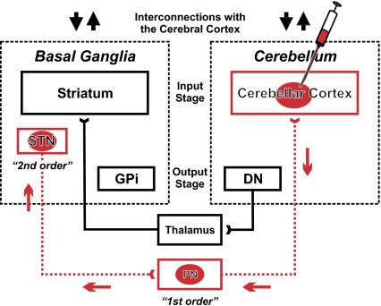
The basal ganglia and cerebellum are major subcortical structures that influence not only movement, but putatively also cognition and affect. Both structures receive input from and send output to the cerebral cortex. Thus, the basal ganglia and cerebellum form multisynaptic loops with the cerebral cortex. Basal ganglia and cerebellar loops have been assumed to be anatomically separate and to perform distinct functional operations. We investigated whether there is any direct route for basal ganglia output to influence cerebellar function that is independent of the cerebral cortex. We injected rabies virus (RV) into selected regions of the cerebellar cortex in cebus monkeys and used retrograde transneuronal transport of the virus to determine the origin of multisynaptic inputs to the injection sites. We found that the subthalamic nucleus of the basal ganglia has a substantial disynaptic projection to the cerebellar cortex. This pathway provides a means for both normal and abnormal signals from the basal ganglia to influence cerebellar function. We previously showed that the dentate nucleus of the cerebellum has a disynaptic projection to an input stage of basal ganglia processing, the striatum. Taken together these results provide the anatomical substrate for substantial two-way communication between the basal ganglia and cerebellum. Thus, the two subcortical structures may be linked together to form an integrated functional network.


Conflict of interest statement

The authors declare no conflict of interest.

Figures

Fig. 1.
Fig. 1.
Experimental paradigm and circuits interconnecting basal ganglia and cerebellum. We injected rabies virus (RV) into regions of the cerebellar hemisphere. The virus went through two stages of transport: retrograde transport to first-order neurons that innervate the injection site and then, retrograde transneuronal transport to second-order neurons that innervate the first-order neurons. The red arrows indicate the direction of virus transport. Previously, we have shown that an output stage of cerebellar processing, the dentate nucleus (DN), has a disynaptic connection with an input stage of basal ganglia processing, the striatum (4). In this experiment, we demonstrate a reciprocal connection from the subthalamic nucleus (STN) to the input stage of cerebellar processing, the cerebellar cortex. These interconnections enable two-way communication between the basal ganglia and cerebellum. Each of these subcortical modules has separate parallel interconnections with the cerebral cortex (up and down black arrows). DN, dentate nucleus; GPi, internal segment of the globus pallidus; PN, pontine nuclei; STN, subthalamic nucleus.
Fig. 2.
Fig. 2.
Injection sites and second-order neurons labeled in STN. (A) The injection sites of rabies virus (RV) with cholera toxin subunit β (CTb) are outlined on a flattened map of the cerebellar cortex adapted from ref. . The injection in AB2 (red filled area) targeted Crus IIp. The injection site in another animal (AB1, not illustrated) also targeted Crus IIp. In this case the injection site overlapped, but was somewhat less extensive than that of AB2. The injection in AB3 (blue filled area) targeted HVIIB. (B) Cross-sections of the STN show the location of second-order neurons labeled by the retrograde transneuronal transport of RV from Crus IIp in AB2 (red dots) and from HVIIB in AB3 (blue dots). Each of the three rostrocaudal levels displayed is spaced ≈1 mm apart. Labeled neurons from three consecutive sections (spaced 100 μm apart) are overlapped at each level. a, anterior; C, caudal; D, dorsal; F.amp., ansoparamedian fissure; F.in.cr., intracrural fissure; F.ppd., prepyramidal fissure; F.pr., primary fissure; F.ps., posterior superior fissure; M, medial; p, posterior.
Fig. 3.
Fig. 3.
Second-order neurons in the STN labeled by the retrograde transneuronal transport of RV. (A) Chart of a coronal section through the midbrain in one monkey (AB2). Each dot represents a neuron infected with RV. (B) Photomicrograph of the boxed area in A. Arrows point to examples of second-order neurons labeled with RV. (C) Enlargement of the boxed area in B. D, dorsal; M, medial; RNpc, parvocellular red nucleus; SNpc, substantia nigra pars compacta; SNr, substantia nigra pars reticulata; ZI, zona incerta.
Fig. 4.
Fig. 4.
Topography of STN projections to the cerebellar hemisphere. (A) Histogram of the rostrocaudal distribution of second-order neurons in STN in AB2 (red bars) and AB3 (blue bars). The distance between two consecutive sections represented is 100 μm. Missing bars correspond to missing sections. (B) Charts of labeled neurons in AB2 (red dots) and AB3 (blue dots) are overlapped to illustrate the topographic differences in distribution of second-order neurons in STN of the two cases. (C) Schematic representation of STN organization, according to the tripartite functional subdivision of the basal ganglia (adapted from ref. 16). (D) Schematic summary of the known connections of the STN with areas of the cerebral cortex (based on refs. –36). C, caudal; D, dorsal; M, medial.

References

    1. Alexander GE, DeLong MR, Strick PL. Parallel organization of functionally segregated circuits linking basal ganglia and cortex. Annu Rev Neurosci. 1986;9:357–381. - PubMed
    1. Strick PL, Dum RP, Fiez JA. Cerebellum and nonmotor function. Annu Rev Neurosci. 2009;32:413–434. - PubMed
    1. Percheron G, François C, Talbi B, Yelnik J, Fénelon G. The primate motor thalamus. Brain Res Brain Res Rev. 1996;22:93–181. - PubMed
    1. Hoshi E, Tremblay L, Féger J, Carras PL, Strick PL. The cerebellum communicates with the basal ganglia. Nat Neurosci. 2005;8:1491–1493. - PubMed
    1. Kelly RM, Strick PL. Rabies as a transneuronal tracer of circuits in the central nervous system. J Neurosci Methods. 2000;103:63–71. - PubMed

Publication types

LinkOut - more resources