. 2010 Jun 11;285(24):18939-47.
doi: 10.1074/jbc.M110.103028. Epub 2010 Apr 19.

Canonical Wnt3a modulates intracellular calcium and enhances excitatory neurotransmission in hippocampal neurons

Affiliations

Canonical Wnt3a modulates intracellular calcium and enhances excitatory neurotransmission in hippocampal neurons

Miguel E Avila et al. J Biol Chem. .

Erratum in

Abstract

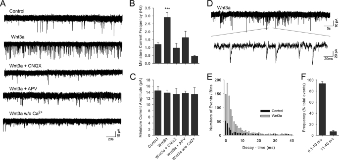
A role for Wnt signal transduction in the development and maintenance of brain structures is widely acknowledged. Recent studies have suggested that Wnt signaling may be essential for synaptic plasticity and neurotransmission. However, the direct effect of a Wnt protein on synaptic transmission had not been demonstrated. Here we show that nanomolar concentrations of purified Wnt3a protein rapidly increase the frequency of miniature excitatory synaptic currents in embryonic rat hippocampal neurons through a mechanism involving a fast influx of calcium from the extracellular space, induction of post-translational modifications on the machinery involved in vesicle exocytosis in the presynaptic terminal leading to spontaneous Ca(2+) transients. Our results identify the Wnt3a protein and a member of its complex receptor at the membrane, the low density lipoprotein receptor-related protein 6 (LRP6) coreceptor, as key molecules in neurotransmission modulation and suggest cross-talk between canonical and Wnt/Ca(2+) signaling in central neurons.


Figures

FIGURE 1.
FIGURE 1.
Purification and functional assays of Wnt3a activity. A, Coomassie Blue staining of an SDS-PAGE gel that was loaded with Wnt3a L-cell conditioned medium (CM) and purified Wnt3a, resulting from a three-step chromatographic purification. BS, Blue Sepharose; HiL, High Load 16–60 Superdex 200; Hep, heparin; MW, molecular mass. B and C, Western blotting analysis of purified Wnt3a and time-dependent effect on β-catenin stabilization in hippocampal neurons following different periods of Wnt3a application (15 min, 30 min, and 2 h); LiCl (10 mm) for 2 h was used as a control. D, summary of data as shown in C. E, β-catenin reporter activity in pBARL-HT22 cells treated with 10 nm Wnt3a, Wnt3a vehicle (buffer), boiled Wnt3a (denatured 10 min at 96 °C), 10 nm Wnt3a plus 50 nm recombinant sFRP1, 50 nm sFRP1, Wnt3a plus 70 nm recombinant DKK1, and 70 nm DKK1. Control, basal reporter activity in this cell line. The data from three independent experiments are shown as the means ± S.E., and test ANOVA (*, p < 0.05; ***, p < 0.005) was implemented.
FIGURE 2.
FIGURE 2.
Enhancement of miniature synaptic activity by purified Wnt3a in primary cultures of hippocampal neurons. A, representative traces of spontaneous synaptic currents recorded in the presence of different Wnt3a concentrations (0–10 nm). B, the upper panel shows representative traces of individual events in voltage records (action potentials) obtained from control neurons and those treated with 10 nm of the Wnt3a protein. The lower panel depicts a repetitive event induced by 10 nm Wnt3a. The dotted lines mark 0 mV. The records (n = 5) were obtained in the absence of TTX, and the treatments were applied by perfusion. C, miniature synaptic activity following the application of Wnt3a (5 and 15 min) and various treatments by perfusion in the presence of 100 nm TTX (holding potential of −60 mV; 2-min duration). D and E, summary of miniature synaptic activity data for current frequency and amplitude, respectively. Control, Wnt3a vehicle; Wnt3a, 10 nm Wnt3a; Boiled, denatured Wnt3a (boiled 96 °C); anti-Wnt3a, antibody anti-Wnt3a; Boiled anti-Wnt3a, boiled antibody anti-Wnt3a (96 °C); washout, Wnt3a removed from external solution. Test ANOVA was implemented (***, p < 0.005; n = 5). The data are shown as the means ± S.E.
FIGURE 3.
FIGURE 3.
Inhibition of Wnt3a-induced synaptic activity by glutamatergic blockers CNQX and APV. A, the miniature synaptic transmission induced by 10 nm Wnt3a (15 min) in hippocampal neurons was pharmacologically blocked following application of CNQX (4 μm) or APV (50 μm) by perfusion in the presence of ligand and TTX (100 nm) and in the absence of Mg2+. B and C, data summary for the effects on the frequency and amplitude of the miniature currents, respectively. Control, Wnt3a vehicle; w/o Ca2+, zero nominal calcium. The data are shown as the means ± S.E., and test ANOVA (*, p < 0.05; ***, p < 0.005; n = 5) was implemented. D–F, analysis of an extended trace of miniature synaptic activity recorded in the presence of 10 nm Wnt3a. E and F, stacked bars plot showing event decay-time (ms) distribution histogram and the frequency of total events in Wnt3a-treated neurons. 0.1–10 ms, fast AMPAergic events; 10–40 ms, slow GABAergic events.
FIGURE 4.
FIGURE 4.
Enhancement of intracellular Ca2+ and synaptic vesicle release by Wnt3a in hippocampal neurons. A, representative fluorescent traces showing spontaneous enhancement of calcium transients following application of Wnt3a and various treatments for 15 min. B, strokes representing the effect of acute influx of extracellular Ca2+ following perfusion of Wnt3a and various treatments. C and D, synaptic vesicle release from presynaptic terminals induced by Wnt3a. C, destaining associated to FM1–43 (depleted fraction, ΔF/Fi) in the presence or absence of Wnt3a. Treatments were applied by perfusion during the entire record (20 min), and the burden of hippocampal neurons was recorded (n = 60–80 neurons). D, immunocytochemical analysis showing the decay of the signal associated with the fixable AM1–43 probe (upper panels) and the enhancement of phosphorylated Synapsin I (Ser-553) after treatment with Wnt3a for 15 min (lower panels). The calibration bar represents 20 μm. E, Western blot of phosphorylated Synapsin I after treatment for 15 min with purified Wnt3a compared with control neurons either treated with boiled Wnt3a or coincubated with a Wnt3a-specific antibody. All records of Ca2+ changes were made in the absence of TTX. Control, Wnt3a vehicle; ENS, external normal solution containing 10 nm Wnt3a; Cd2+, ENS plus 10 nm Wnt3a and 20 μm Cd2+; w/o Ca2+, 10 nm Wnt3a plus external solution without Ca2+ (zero nominal Ca2+); Boiled, denatured Wnt3a (boiled 10 min to 96 °C); anti-Wnt3a, coincubation of 10 nm Wnt3a and 2 μg/ml of antibody anti-Wnt3a.
FIGURE 5.
FIGURE 5.
Evidence for the participation of the Wnt/β-catenin complex receptor in Wnt3a-induced neurotransmission. A and B, representative traces showing the inhibition of the Wnt3a enhancement in the frequency of miniature synaptic activity (test ANOVA; ***, p < 0.005; n = 5; 100 nm TTX) and calcium transients (test ANOVA; ***, p < 0.005; n = 40–50), respectively, following coincubation for 15 min of 10 nm Wnt3a with either 50 nm sFRP1 or 70 nm DKK1, which act as inhibitors of the Wnt/β-catenin membrane-associated receptors (see also Fig. 1E). C and D, summary of miniature synaptic activity frequency and calcium transient data, respectively. The data are shown as the means ± S.E.
FIGURE 6.
FIGURE 6.
LRP6 immunoreactivity in the soma and neuronal processes associated with pre- and postsynaptic markers Syp and PSD95. A, confocal images were obtained from neurons of 14–15 DIV using antibodies against LRP6 (green, Alexa488), Syp (red, Cy3), and PSD95 (blue, Alexa633). The arrows show LRP6-Syp and LRP6-PSD95 colocalization. Calibration bars, 20 μm. B and C, Mander's and Pearson's correlation coefficients for the colocalization of the LRP6 and pre- and postsynaptic markers Syp and PSD95, respectively. The averages of coefficients of at least three regions of interest of 10 neurons were examined.

References

    1. Grove E. A., Tole S., Limon J., Yip L., Ragsdale C. W. (1998) Development 125, 2315–2325 - PubMed
    1. Houart C., Caneparo L., Heisenberg C., Barth K., Take-Uchi M., Wilson S. (2002) Neuron 35, 255–265 - PubMed
    1. Lee S. M., Tole S., Grove E., McMahon A. P. (2000) Development 127, 457–467 - PubMed
    1. Maretto S., Cordenonsi M., Dupont S., Braghetta P., Broccoli V., Hassan A. B., Volpin D., Bressan G. M., Piccolo S. (2003) Proc. Natl. Acad. Sci. U.S.A. 100, 3299–3304 - PMC - PubMed
    1. Zhou C. J., Zhao C., Pleasure S. J. (2004) J. Neurosci. 24, 121–126 - PMC - PubMed

Publication types