. 2010 Jun 18;285(25):19277-87.
doi: 10.1074/jbc.M109.079939. Epub 2010 Apr 19.

Amelogenin-collagen interactions regulate calcium phosphate mineralization in vitro

Affiliations

Amelogenin-collagen interactions regulate calcium phosphate mineralization in vitro

Atul S Deshpande et al. J Biol Chem. .

Abstract

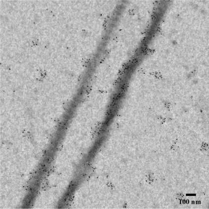
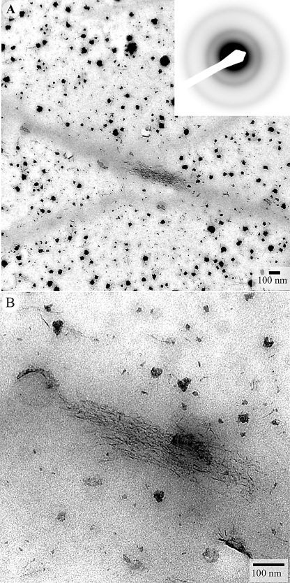
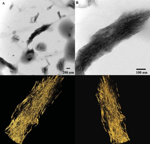
Collagen and amelogenin are two major extracellular organic matrix proteins of dentin and enamel, the mineralized tissues comprising a tooth crown. They both are present at the dentin-enamel boundary (DEB), a remarkably robust interface holding dentin and enamel together. It is believed that interactions of dentin and enamel protein assemblies regulate growth and structural organization of mineral crystals at the DEB, leading to a continuum at the molecular level between dentin and enamel organic and mineral phases. To gain insight into the mechanisms of the DEB formation and structural basis of its mechanical resiliency we have studied the interactions between collagen fibrils, amelogenin assemblies, and forming mineral in vitro, using electron microscopy. Our data indicate that collagen fibrils guide assembly of amelogenin into elongated chain or filament-like structures oriented along the long axes of the fibrils. We also show that the interactions between collagen fibrils and amelogenin-calcium phosphate mineral complexes lead to oriented deposition of elongated amorphous mineral particles along the fibril axes, triggering mineralization of the bulk of collagen fibril. The resulting structure was similar to the mineralized collagen fibrils found at the DEB, with arrays of smaller well organized crystals inside the collagen fibrils and bundles of larger crystals on the outside of the fibrils. These data suggest that interactions between collagen and amelogenin might play an important role in the formation of the DEB providing structural continuity between dentin and enamel.


Figures

FIGURE 1.
FIGURE 1.
TEM micrographs of collagen fibrils incubated with amelogenin and negatively stained, at low (A) and intermediate (B) magnifications. Note the chains of nanospheres and fused filamentous amelogenin assemblies associated with collagen fibrils, indicated by arrowheads.
FIGURE 2.
FIGURE 2.
TEM micrographs of collagen fibrils incubated with amelogenin and labeled with amelogenin antibodies.
FIGURE 3.
FIGURE 3.
TEM micrographs of unstained collagen fibrils mineralized in the presence of amelogenin for 2 h, at low (A) and intermediate (B) magnifications. The diffraction pattern in the inset in panel A suggests the presence of ACP. In panel B a close up of the outlined mineral aggregate in the inset, arrowheads point toward individual mineral particles forming the mineral aggregate. Panel C shows four representative high magnification images of filamentous mineral particles (arrowheads). Micrograph in D shows the mineral particles surrounded by low electron density protein envelopes (arrows).
FIGURE 4.
FIGURE 4.
TEM micrographs of unstained collagen fibrils mineralized in the presence of amelogenin for 3 h, at low (A) and high magnifications (B). The diffraction pattern in the inset suggests the presence of ACP. Note the bundles of filamentous mineral on the collagen fibrils surfaces aligned with the fibril axes. Note also that elongated mineral particles similar to those forming on the collagen fibrils are distributed throughout the field of view, however, they are not organized.
FIGURE 5.
FIGURE 5.
TEM micrographs of unstained collagen fibrils mineralized in the presence of amelogenin for 4 h at low (A) and intermediate (B); C is a higher magnification of the fibril in B. The mineral is poorly crystalline apatitic phase, based on the electron diffraction in the inset, which is taken from the mineralize collagen fibril (f) shown in C. Note that the 002 reflections are co-aligned with the collagen fibril axis. Also note small bundles of elongated crystals throughout the grid (arrows).
FIGURE 6.
FIGURE 6.
TEM micrographs of unstained collagen fibrils mineralized in the presence of amelogenin for 16 h. Electron diffraction pattern in the inset taken from the fibril in A, indicates the presence of mature apatitic crystals co-aligned with the fibril axis. Besides the mineral particles associated with collagen fibrils, bundles of elongated crystals (arrowheads) were observed throughout the grid (B).
FIGURE 7.
FIGURE 7.
A, TEM micrograph of a collagen fibril with mineral bundles attached at its surface. B, a tomographic reconstruction of the area outlined in A. 1 and 2 are views from different angles at mineralized bundles indicated in B as areas 1 and 2.
FIGURE 8.
FIGURE 8.
A, TEM micrographs of fully mineralized collagen fibril at low (A) and high magnifications (B). Three-dimensional reconstructions of the mineralized fibril from two different angles are shown in the bottom panel.

Similar articles

Cited by

References

    1. Nanci A. (2007) Ten Cate's Oral Histology: Development, Structure, and Function, Mosby, St. Louis, MO
    1. Weiner S., Wagner H. D. (1998) Annu. Rev. Materials Sci. 28, 271–298
    1. Landis W. J., Hodgens K. J., Arena J., Song M. J., McEwen B. F. (1996) Microsc. Res. Tech. 33, 192–202 - PubMed
    1. Weiner S., Traub W. (1992) FASEB J. 6, 879–885 - PubMed
    1. Brodsky B., Ramshaw J. A. M. (1997) Matrix Biol. 15, 545–554 - PubMed

Publication types

LinkOut - more resources