. 2010 Jun 18;285(25):19450-9.
doi: 10.1074/jbc.M110.109793. Epub 2010 Apr 19.

Mzm1 influences a labile pool of mitochondrial zinc important for respiratory function

Affiliations

Mzm1 influences a labile pool of mitochondrial zinc important for respiratory function

Aaron Atkinson et al. J Biol Chem. .

Abstract

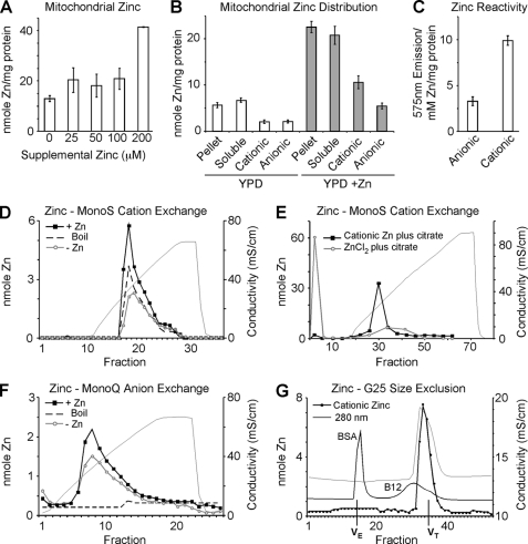
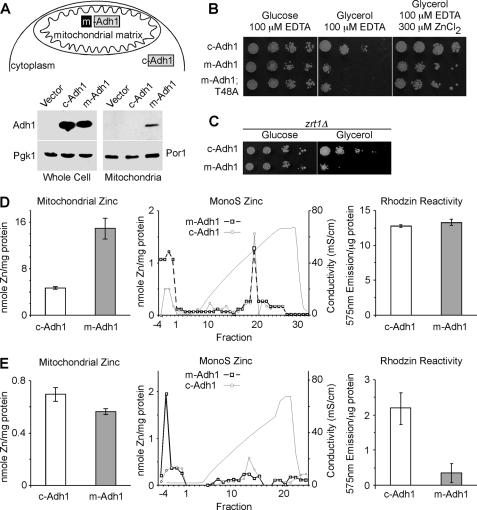
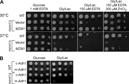
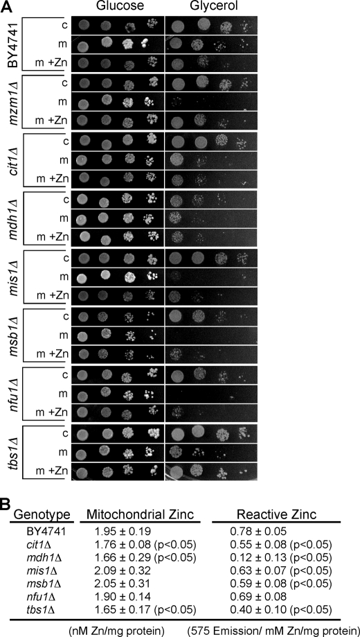
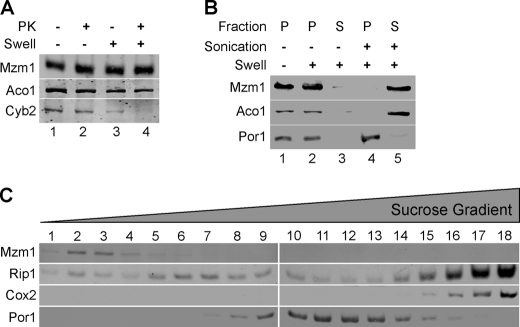
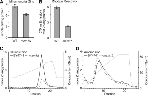
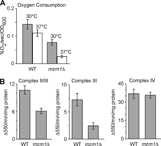
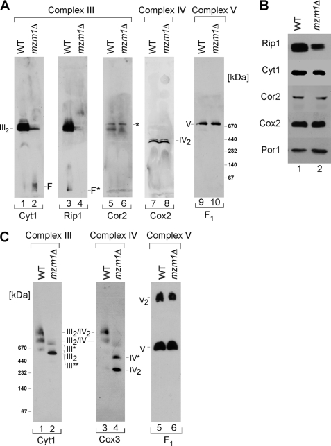
Zinc is essential for function of mitochondria as a cofactor for several matrix zinc metalloproteins. We demonstrate that a labile cationic zinc component of low molecular mass exists in the yeast mitochondrial matrix. This zinc pool is homeostatically regulated in response to the cellular zinc status. This pool of zinc is functionally important because matrix targeting of a cytosolic zinc-binding protein reduces the level of labile zinc and interferes with mitochondrial respiratory function. We identified a series of proteins that modulate the matrix zinc pool, one of which is a novel conserved mitochondrial protein designated Mzm1. Mutant mzm1Delta cells have reduced total and labile mitochondrial zinc, and these cells are hypersensitive to perturbations of the labile pool. In addition, mzm1Delta cells have a destabilized cytochrome c reductase (Complex III) without any effects on Complexes IV or V. Thus, we have established that a link exists between Complex III integrity and the labile mitochondrial zinc pool.


Figures

FIGURE 1.
FIGURE 1.
Mitochondrial zinc pools. Yeast cultures were grown in yeast peptone 1% dextrose (YP1D) medium and supplemented with 0–200 μm ZnCl2. Mitochondria were isolated from cultures in triplicate and analyzed for zinc either from total or submitochondrial fractions by ICP-OES. Panel A, titration of supplemental zinc to cell cultures results in expansion of mitochondrial zinc. Panel B, soluble and pellet fractions were isolated from 0.5 mg of mitochondria by sonication and centrifugation. Ion exchange using either CM-52 or DEAE-52 cellulose was used to separate cationic and anionic fractions respectively. Panel C, using the Zn-dependent fluorophore Rhodzin-3, reactive zinc was assessed in mitochondrial lysate from 0.1 mg of mitochondria depleted of either anionic or cationic Zn pools (post DEAE-52 or CM-52 binding, respectively; n = 4). Panels D–F, MonoS and MonoQ ion exchange chromatography. Mitochondria were isolated from YP1D yeast cultures grown with 0.2 mm Zn (+Zn) or without (−Zn, Endogenous) and samples (equivalent to 0.5 mg of protein) were sonicated, clarified, and boiled where indicated. Panel D, fractionation of soluble lysate using MonoS shows a single cationic zinc peak that expands in response to zinc supplementation and is resistant to boiling. Panel E, either free Zn(II) in the form of ZnCl2 or previously purified mitochondrial cationic Zn were mixed with a 10-fold excess of citrate and examined for the ability to bind to MonoS cation exchange resin. Panel F, fractionation of soluble lysate using MonoQ anion exchange reveals a broad peak that expands modestly in response to zinc supplementation, yet is not resistant to boiling. Panel G, gel filtration of the cationic zinc fraction isolated by MonoS elutes on a G25 size exclusion column in a volume corresponding to salt. 280 nm absorbance for both bovine serum albumin (BSA) and Vitamin B12 (B12) are shown. Volume exclusion (VE) and volume total (VT) are marked.
FIGURE 2.
FIGURE 2.
Effect of matrix-targeted Adh1 (m-Adh1) on mitochondrial zinc. Upper panel A, a schematic depicting the N-terminal fusion of the Sod2 mitochondrial targeting sequence directing Adh1 to the matrix, where upon import the Sod2 presequence is removed. Lower panel A, relative expression of c-Adh1 and m-Adh1 in both whole cell extracts and gradient-purified mitochondria relative to the respective loading controls Pgk1 and Por1. Panel B, wild-type yeast expressing either m-Adh1, or a catalytic inactive m-Adh1 mutant (T48A), show respiratory phenotypes when grown under Zn-limited conditions that are suppressed in the presence of excess Zn. Panel C, yeast lacking the plasma membrane zinc transporter, Zrt1, show an m-Adh1-induced respiratory phenotype without EDTA supplementation. Left panel D, total mitochondrial zinc from both c-Adh1- and m-Adh1-expressing cells obtained from cultures grown in synthetic complete Zn-replete medium. Right panel D, MonoS fractionation and Rhodzin-3 fluorescence (n = 3) of soluble mitochondrial lysates from either c-Adh1- or m-Adh1-expressing cultures. Left panel E, total mitochondrial zinc from both c-Adh1- and m-Adh1-expressing cells obtained from cultures grown in synthetic complete Zn-deficient medium. Right panel E, MonoS fractionation and Rhodzin-3 fluorescence (n = 3) of soluble mitochondrial lysates Zn-deficient cultures.
FIGURE 3.
FIGURE 3.
Zn-dependent respiratory defects. In a large-scale screen for mutants showing Zn-dependent respiratory defects, we isolated mzm1Δ from a collection of 684 deletion strains lacking proteins predicted to have mitochondrial localization. Panel A, mzm1Δ cells are unable to grow on respiratory carbon sources under Zn-limited conditions (EDTA) or at elevated temperature relative to both wild type and the complemented strain. Panel B, the presence of m-Adh1 severely restricts mzm1Δ respiratory growth.
FIGURE 4.
FIGURE 4.
m-Adh1-induced respiratory deficiency. Panel A, yeast mzm1Δ, cit1Δ, mdh1Δ, mis1Δ, msb1Δ, nfu1Δ, and tbs1Δ strains were identified as m-Adh1 (T48A) sensitive from the collection of 684 strains lacking predicted mitochondrial proteins. Strains were re-transformed with either c-Adh1 or m-Adh1 (T48A) and grown in either synthetic complete (SC) glucose medium without (−Zn) or with (+Zn) 500 μm supplemental ZnCl2. Cultures were then serially diluted onto SC medium agar with glucose or glycerol as carbon sources. All strains were verified by PCR using a universal KanMX and gene-specific primers. Panel B, mitochondria from cit1Δ, mdh1Δ, mis1Δ, msb1Δ, nfu1Δ, and tbs1Δ cells were purified from cells grown in standard SC 1% glucose medium for comparison to wild-type mitochondria. Both total mitochondrial zinc and reactive zinc, as assessed with Rhodzin-3, were measured in triplicate on three independent mitochondrial preparations.
FIGURE 5.
FIGURE 5.
Localization of Mzm1. Panel A, mitochondria (100 μg) expressing Mzm1-Myc were kept intact (− swell, lanes 1 and 2) or swollen (+ swell, lanes 3 and 4) and incubated with (lanes 2 and 4) or without (lanes 1 and 3) proteinase K (PK). The matrix marker protein is Aco1, mitochondrial aconitase; inner mitochondrial space protein is Cyb2, cytochrome b2. Panel B, mitochondria (100 μg of protein) were fractionated using both hypotonic lysis (swelling) and sonication. Pellet (P) and soluble (S) fractions were isolated by centrifugation (see “Materials and Methods” for details). In swollen mitoplasts in the absence of sonication, Mzm1 and Aco1 are retained in the pellet fraction. Detection with porin (Por1) antibody marks the pellet fraction. Panel C, mitochondria (1 mg) were solubilized in buffer containing 1% digitonin and centrifuged over a continuous 7–30% sucrose gradient. Mzm1-Myc migration is shown relative to Por1 and markers for Complex III (Rip1), and for Complex IV (Cox2).
FIGURE 6.
FIGURE 6.
Mitochondrial Zn defect in mzm1Δ cells. Mitochondria were isolated for both WT and mzm1Δ from cells grown in yeast peptone 1% dextrose. Panel A, metal analysis of mitochondria (200 μg; n = 3). Panel B, mitochondria (50 μg) were sonicated in 0.1 ml 10 mm Tris, and the clarified lysate incubated with Rhodzin-3 to asses Zn-dependent fluorescence. Following fluorescent measurements, lysates were analyzed for metal content by ICP-OES (n = 3). Panel C, MonoS fractionation of mzm1Δ soluble mitochondrial lysate. Panel D, MonoQ fractionation of mzm1Δ soluble mitochondrial lysate.
FIGURE 7.
FIGURE 7.
Respiratory defect in mzm1Δ cells. Panel A, total oxygen consumption of mzm1Δ relative to wild type at 30° and 37 °C. Panel B, analysis of succinate cytochrome c reductase activity (Complex II/III), ubiquinol cytochrome c reductase activity (Complex III), and cytochrome c oxidase activity (Complex IV) using 10–20 μg of mitochondria per reaction (see “Materials and Methods” for details) (n = 3 per assay).
FIGURE 8.
FIGURE 8.
The bc1 complex is impaired in mzm1Δ cells. Panel A, mitochondria (75 μg) were solubilized in buffer containing 1% dodecyl-β-d-maltoside (DDM) and analyzed by BN-PAGE. Detection with Cyt1 (cytochrome c1, lanes 1 and 2), Rip1 (Rieske Iron-sulfur Protein 1, lanes 3 and 4), and Cor2 (core protein 2, lanes 5 and 6) reveal the state of dimeric Complex III (III2) in both WT and mzm1Δ. Detection of either Cox2 (lanes 7 and 8) or F1 (lanes 9 and 10) representing Complex IV and V, respectively, shows no defects between mzm1Δ and WT cells. Low molecular weight or free Cyt1 (F); low molecular weight or free Rip1 (F*); residual Complex V signal (*). Panel B, immunoblot analysis of wild-type and mzm1Δ cells for protein levels of Complex III subunits Rip1, Cyt1, and Cor2 as well as Cox2 (Complex IV) and porin. Panel C, mitochondria (150 μg) solubilized in buffer containing 1% digitonin were fractionated on a continuous 5–13% gradient gel and protein analyzed by BN-PAGE. Detection with the Complex III subunit Cyt1 reveals subassembly complexes (III*, and III**) together with dimeric Complex III (III2) and the higher Complex III/IV supercomplexes (III2/IV, and III2/IV2). Detection with the Complex IV component, Cox3, also reveals a Complex IV intermediate (IV*) in addition to dimeric Complex IV (IV2). Detection with the Complex V subunit, F1, reveals no defect between mzm1Δ and wild type.

Similar articles

Cited by

References

    1. Atkinson A., Winge D. R. (2009) Chem. Rev. 109, 4708–4721 - PMC - PubMed
    1. Mesecke N., Bihlmaier K., Grumbt B., Longen S., Terziyska N., Hell K., Herrmann J. M. (2008) EMBO Rep. 9, 1107–1113 - PMC - PubMed
    1. Coyne H. J., 3rd, Ciofi-Baffoni S., Banci L., Bertini I., Zhang L., George G. N., Winge D. R. (2007) J. Biol. Chem. 282, 8926–8934 - PubMed
    1. Eide D. J. (2006) Biochim. Biophys. Acta 1763, 711–722 - PubMed
    1. Jackson K. A., Helston R. M., McKay J. A., O'Neill E. D., Mathers J. C., Ford D. (2007) J. Biol. Chem. 282, 10423–10431 - PubMed

Publication types