. 2010 Jul 2;285(27):21114-25.
doi: 10.1074/jbc.M109.096479. Epub 2010 Apr 19.

Versican facilitates chondrocyte differentiation and regulates joint morphogenesis

Affiliations

Versican facilitates chondrocyte differentiation and regulates joint morphogenesis

Kanyamas Choocheep et al. J Biol Chem. .

Abstract

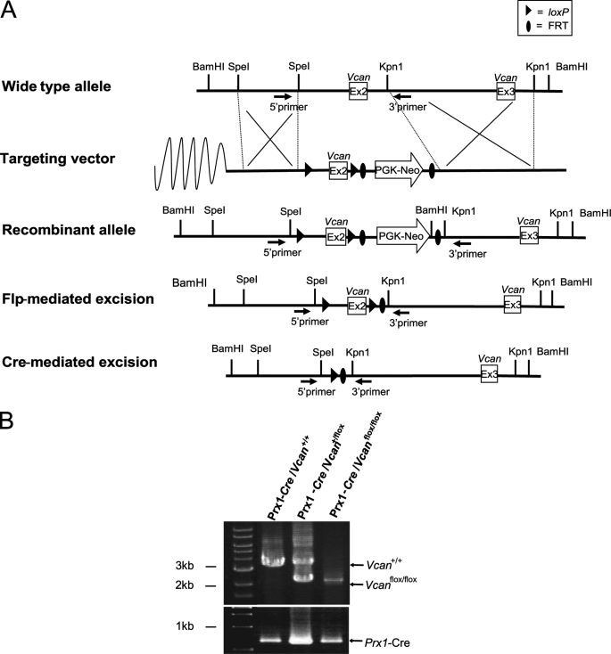
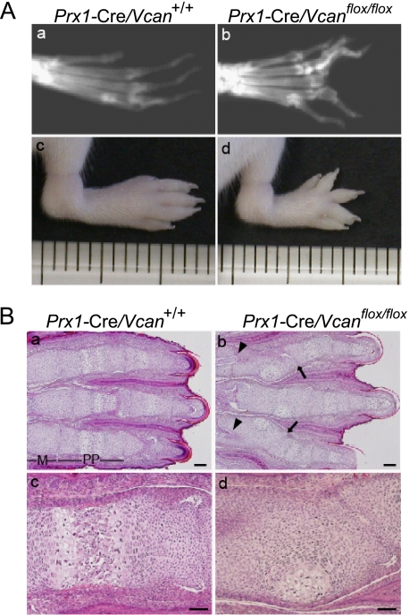
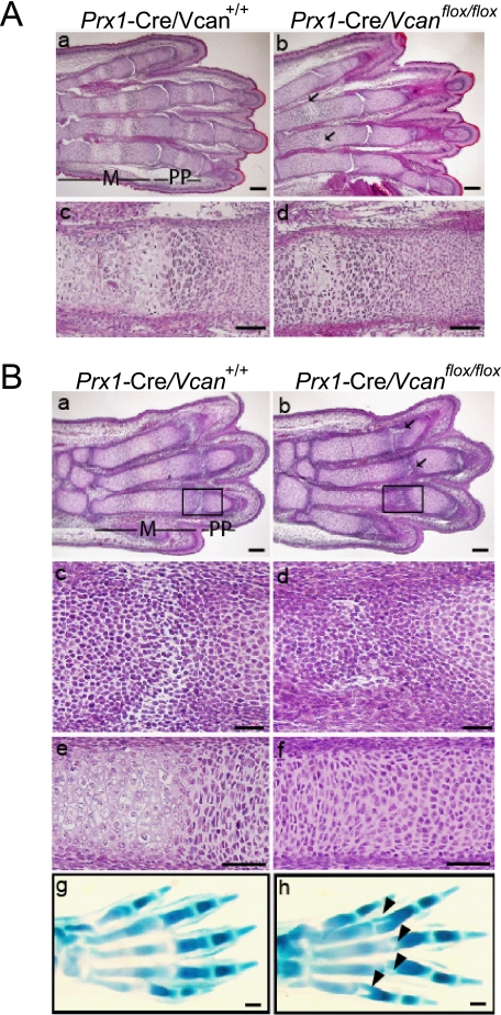
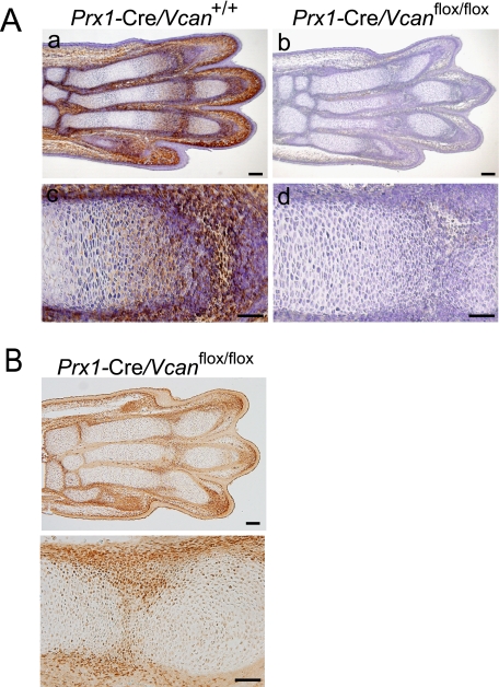
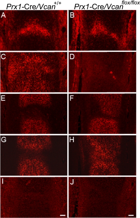
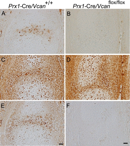
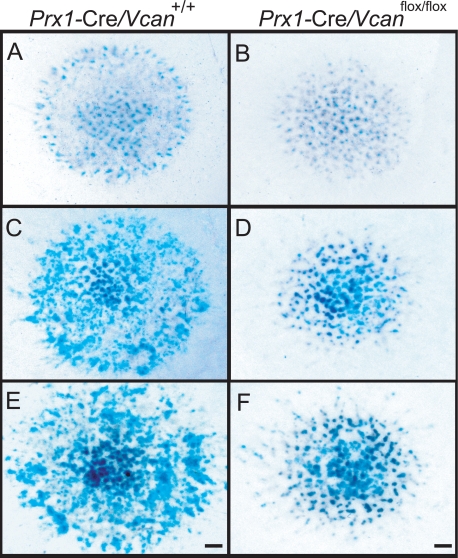
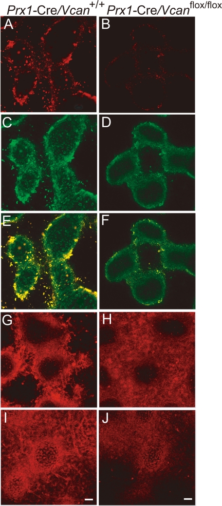
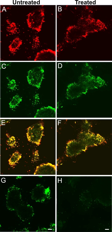
Versican/PG-M is a large chondroitin sulfate proteoglycan in the extracellular matrix, which is transiently expressed in mesenchymal condensation areas during tissue morphogenesis. Here, we generated versican conditional knock-out mice Prx1-Cre/Vcan(flox/flox), in which Vcan is pruned out by site-specific Cre recombinase driven by the Prx1 promoter. Although Prx1-Cre/Vcan(flox/flox) mice are viable and fertile, they develop distorted digits. Histological analysis of newborn mice reveals hypertrophic chondrocytic nodules in cartilage, tilting of the joint, and a slight delay of chondrocyte differentiation in digits. By immunostaining, whereas the joint interzone of Prx1-Cre/Vcan(+/+) shows an accumulation of TGF-beta, concomitant with versican, that of Prx1-Cre/Vcan(flox/flox) without versican expression exhibits a decreased incorporation of TGF-beta. In a micromass culture system of mesenchymal cells from limb bud, whereas TGF-beta and versican are co-localized in the perinodular regions of developing cartilage in Prx1-Cre/Vcan(+/+), TGF-beta is widely distributed in Prx1-Cre/Vcan(flox/flox). These results suggest that versican facilitates chondrogenesis and joint morphogenesis, by localizing TGF-beta in the extracellular matrix and regulating its signaling.


Figures

FIGURE 1.
FIGURE 1.
Genomic construct for conditional deletion of the Vcan mice. A, the wild-type Vcan locus is depicted on the top line. A Vcan targeting vector was constructed by flanking exon 2 with loxP sites, and flanking a PGK-neoR cassette with FRT sites (Targeting vector). ES cell clones with homologous recombination of the vector segment (Recombinant allele) were obtained by positive selection and used for generation of chimera mice, followed by germ line transmission. The FRT-flanked PGK-neoR cassette was subsequently deleted from the recombinant allele by crossing with CAG-Flp Tg mice. These mice, termed Vcanflox/flox, were crossed with Prx1-Cre Tg mice. These mice have cells with the null allele of Vcan, in regions where Prx1 promoter is and was active. B, genotyping was performed by PCR analysis on DNA prepared from a tail biopsy using the primers shown by black arrows. The WT (Vcan+/+) PCR product is 3 kb, and the flox (Vcanflox/flox) product without PGK-neoR cassette is 2 kb. The Prx1-Cre transgene was detected by PCR using another primer set. The reaction product (∼620 bp) is shown.
FIGURE 2.
FIGURE 2.
Conditional removal of Vcan in limb bud mesenchyme leads to limb deformities. A, x-ray visualization of 8-month-old mice (a and b) and gross observations of 1-week-old mouse hind limbs (c and d). Prx1-Cre/ Vcanflox/flox mice (b and d) show distortion and shortening of digits, compared with Prx1-Cre/Vcan+/+ mice (a and c). B, histological analysis of newborn Prx1-Cre/Vcan+/+ (a and c) and Prx1-Cre/Vcanflox/flox (b and d) hind limbs by hematoxylin and eosin staining. Prx1-Cre/Vcanflox/flox digits display joint tilting (arrows in b) and clefting (arrowheads in b), formation of hypertrophic chondrocyte nodules in proximal phalanges, and delayed endochondral ossification (d). M, metatarsus; pp, proximal phalanges. Scale bars, 200 μm (a and b) and 40 μm (c and d). All of the histological sections (n ≥ 5) of Prx1-Cre/Vcanflox/flox digits show similar abnormalities.
FIGURE 3.
FIGURE 3.
Prx1-Cre/Vcanflox/flox digits exhibit tilted joint and delayed cartilage development. A, histological analysis of E18.5 Prx1-Cre/Vcan+/+ (a and c) and Prx1-Cre/Vcanflox/flox (b and d) hind limbs by hematoxylin and eosin staining. Prx1-Cre/Vcan+/+ digits demonstrate a well organized columnar structure of prehypertrophic chondrocytes with vascular invasion in metatarsus (m) and proximal phalanges (pp), whereas Prx1-Cre/Vcanflox/flox digits exhibit formation of hypertrophic chondrocyte nodules in a concentric pattern in proximal phalanges (pp) (arrows in b) and delayed endochondral ossification of metatarsus (d). In addition, clefting of some metatarsophalangeal joints is observed (b). Scale bars, 100 μm (a and b) and 40 μm (c and d). B, histological analysis of E15.5 Prx1-Cre/Vcan+/+ and Prx1-Cre/Vcanflox/flox hind limbs by hematoxylin/eosin and Alcian blue staining. Prx1-Cre/Vcan+/+ digits display horizontal stripes of the metatarsophalangeal joint interzones (a and c (as an enlarged image of the boxed area in a)). Prx1-Cre/Vcan+/+ metatarsus shows prehypertrophic and hypertrophic chondrocyte layers (e). In contrast, Prx1-Cre/Vcanflox/flox digits display tilted joint interzones (b and d (as an enlarged image of the boxed area in b)). Prx1-Cre/Vcanflox/flox metatarsus shows prehypertrophic chondrocytes but not hypertrophic chondrocytes (f). Alcian blue staining of Prx1-Cre/Vcan+/+ hind limb shows well aligned stripes of the interzone (g), whereas that of Prx1-Cre/Vcanflox/flox digits reveals tilting of the joint interzone (indicated by arrowheads in h). Scale bars, 150 μm (a and b), 30 μm (c and d), 50 μm (e and f), and 70 μm (g and h). The number of embryos analyzed was as follows: n = 2 each at E18.5; n = 3 and 5 for Prx1-Cre/Vcanflox/flox and Prx1-Cre/Vcan+/+ at E15.5, respectively). At least two paraffin-embedded blocks were obtained, and their tissue sections were used for histological analysis.
FIGURE 4.
FIGURE 4.
Versican is absent in Prx1-Cre/Vcanflox/flox digits. A, immunostaining for versican of E15.5 Prx1-Cre/Vcan+/+ and Prx1-Cre/Vcanflox/flox hind limbs. In Prx1-Cre/Vcan+/+ digits, versican is strongly immunostained in the interzone and perichondrium and moderately immunostained in the proliferative zone of cartilage (a and c), whereas in Prx1-Cre/Vcanflox/flox digits, versican is immunostained neither in the interzone, the perichondrium, nor the proliferative zone of cartilage (b and d). Scale bars, 120 μm (a and b) and 70 μm (c and d). B, immunostaining for Cre enzyme of E15.5 Prx1-Cre/Vcanflox/flox hind limbs at low (top) and high (bottom) magnifications. Cre enzyme is immunostained in the perichondrium and in the tilted interzone. Scale bars, 100 and 50 μm, respectively. Two individual Prx1-Cre/Vcanflox/flox digits showed the same abnormalities.
FIGURE 5.
FIGURE 5.
Distribution of hyaluronan in the joint interzone is unaffected by the absence of versican. Distributions of HA and HA-binding molecules in the metatarsophalangeal joint interzone at E15.5 of Prx1-Cre/Vcan+/+ (A, C, E, G, and I) and Prx1-Cre/Vcanflox/flox (B, D, F, H, and J) are shown. By detection of HA using b-HABP, unincorporated HA is accumulated at a similar level in the joint interzone of both Prx1-Cre/Vcan+/+ and Prx1-Cre/Vcanflox/flox (A and B). Immunofluorescent staining confirms the presence of versican in the interzone, future articular surface, and perichondrium in Prx1-Cre/Vcan+/+ and its absence in Prx1-Cre/Vcanflox/flox (C and D). Immunofluorescent staining for aggrecan, demarcating the cartilage tissue, demonstrates tilting of the joint surface in Prx1-Cre/Vcanflox/flox (E and F). Link protein (LP) is immunostained in cartilage of Prx1-Cre/Vcan+/+ (G), whereas it is immunostained in both cartilage and the joint interzone of Prx1-Cre/Vcanflox/flox (H). CD44 is not detected in both Prx1-Cre/Vcan+/+ and Prx1-Cre/Vcanflox/flox at this stage (I, J), eliminating the possibility of its participation in joint cavitation. Scale bars: 30 μm. Immunostaining was performed on at least two individual digits for Prx1-Cre/Vcan+/+ and Prx1-Cre/Vcanflox/flox, with the same immunostaining patterns.
FIGURE 6.
FIGURE 6.
TGF-β signaling is attenuated in Prx1-Cre/Vcanflox/flox joint interzone. Immunostaining patterns of TGF-β (A and B), TβRII (C and D), and phospho-Smad2/3 (E and F) in the metatarsophalangeal joint interzone at E15.5 of Prx1-Cre/Vcan+/+ (A, C, and E) and Prx1-Cre/Vcanflox/flox (B, D, and F) are shown. Whereas TGF-β is localized in the Prx1-Cre/Vcan+/+ joint interzone (A), it is not detected in Prx1-Cre/Vcanflox/flox (B). TβRII is broadly immunostained in the joint interzone, perichondrium, and chondrocytes of both Prx1-Cre/Vcan+/+ (C) and Prx1-Cre/Vcanflox/flox (D). Whereas phospho-Smad2/3 is found in the nuclei of cells in the Prx1-Cre/Vcan+/+ interzone, it is not detected in Prx1-Cre/Vcanflox/flox. Scale bars, 20 μm. Immunostaining was performed on at least two individual digits for Prx1-Cre/Vcan+/+ and Prx1-Cre/Vcanflox/flox, with the same immunostaining patterns.
FIGURE 7.
FIGURE 7.
Prx1-Cre/Vcanflox/flox exhibits delayed chondrocyte differentiation in micromass culture. Patterns of micromass stained by Alcian blue at day 3 (A and B), 6 (C and D), and 9 (E and F) of Prx1-Cre/Vcan+/+ (A, C, and E) and Prx1-Cre/Vcanflox/flox (B, D, and F) are shown. At day 3, micromass of Prx1-Cre/Vcan+/+ contains some cartilaginous nodules stained with Alcian blue (A), whereas that of Prx1-Cre/Vcanflox/flox contains a smaller number of them (B). At day 6, the number of the nodules positive for Alcian blue in micromass of Prx1-Cre/Vcanflox/flox increases, but it remains smaller than that of Prx1-Cre/Vcan+/+ (C and D). At day 9, the number of cartilaginous nodules in micromass of Prx1-Cre/Vcan+/+ further increases, and the nodules in the center become strongly stained with Alcian blue (E). Although the number of cartilaginous nodules in micromass of Prx1-Cre/Vcanflox/flox increases during culture (F), it remains smaller than Prx1-Cre/Vcan+/+. Scale bars, 300 μm.
FIGURE 8.
FIGURE 8.
TGF-β signaling is altered in the micromass of Prx1-Cre/ Vcanflox/flox. Immunofluorescent staining at day 6 of culture, for versican (A and B), TGF-β (C and D), both merged (E and F), TβRII (G and H), and phospho-Smad2/3 (I and J) of Prx1-Cre/Vcan+/+ (A, C, E, G, and I) and Prx1-Cre/ Vcanflox/flox (B, D, F, H, and J) are shown. Versican is immunostained strongly in the perinodular region and moderately in the internodular region in Prx1-Cre/Vcan+/+ (A), whereas it is immunostained very faintly in Prx1-Cre/Vcanflox/flox (B). TGF-β is mainly localized in the perinodular regions in Prx1-Cre/Vcan+/+ similar to versican (C), whereas it is immunostained diffusely at a substantially lower intensity in Prx1-Cre/Vcan+/+ (D). When merged, both verican and TGF-β are well co-localized in the perinodular region (E). TβRII is immunostained mainly in the perinodular region in both Prx1-Cre/Vcan+/+ (G) and Prx1-Cre/Vcanflox/flox (H) micromass. In addition, it is immunostained rather diffusely in Prx1-Cre/Vcanflox/flox (H). Phospho-Smad2/3 is immunostained in the nuclei of perinodular cells strongly and in chondrocytes in the nodules moderately in Prx1-Cre/Vcan+/+ micromass (I). In contrast, it is immunostained moderately in the nuclei of chondrocytes in the nodules and weakly in the perinodular cells in the Prx1-Cre/Vcanflox/flox micromass (J). Immunofluorescent staining was visualized by Alexa Fluor 594. Scale bars, 13 μm. Immunofluorescent staining was performed at least twice, with essentially the same results. Immunostaining was performed at least three times from individual micromass for Prx1-Cre/Vcan+/+ and Prx1-Cre/Vcanflox/flox, with essentially the same immunostaining patterns.
FIGURE 9.
FIGURE 9.
Versican localizes TGF-β in the extracellular matrix. Immunofluorescent staining of versican (A and B) and TGF-β (C and D) of Prx1-Cre/Vcan+/+ micromass untreated (A, C, and E) and treated with chondroitinase ABC for 48 h before fixation (B, D, and F). The areas immunostained for versican become diffuse by the treatment, although versican essentially remains in the perinodular region (B). The areas immunostained for TGF-β become diffuse by the treatment, concomitantly with versican. Merged images are shown (E and F). Immunostaining for CS chains that confirm ablation of CS is shown (untreated (G) and treated (H)). Scale bars, 15 μm. Immunofluorescent staining was performed at least three times, with essentially the same results.

Similar articles

Cited by

References

    1. Kimata K., Oike Y., Tani K., Shinomura T., Yamagata M., Uritani M., Suzuki S. (1986) J. Biol. Chem. 261, 13517–13525 - PubMed
    1. Matsumoto K., Shionyu M., Go M., Shimizu K., Shinomura T., Kimata K., Watanabe H. (2003) J. Biol. Chem. 278, 41205–41212 - PubMed
    1. Isogai Z., Aspberg A., Keene D. R., Ono R. N., Reinhardt D. P., Sakai L. Y. (2002) J. Biol. Chem. 277, 4565–4572 - PubMed
    1. Aspberg A., Miura R., Bourdoulous S., Shimonaka M., Heinegârd D., Schachner M., Ruoslahti E., Yamaguchi Y. (1997) Proc. Natl. Acad. Sci. U.S.A. 94, 10116–10121 - PMC - PubMed
    1. Olin A. I., Mörgelin M., Sasaki T., Timpl R., Heinegård D., Aspberg A. (2001) J. Biol. Chem. 276, 1253–1261 - PubMed

Publication types

MeSH terms

LinkOut - more resources