Adult plasticity of spatiotemporal receptive fields of multisensory superior colliculus neurons following early visual deprivation
- PMID: 20404413
- PMCID: PMC3652394
- DOI: 10.3233/RNN-2010-0488
Adult plasticity of spatiotemporal receptive fields of multisensory superior colliculus neurons following early visual deprivation
Abstract
Purpose: Previous work has established that the integrative capacity of multisensory neurons in the superior colliculus (SC) matures over a protracted period of postnatal life (Wallace and Stein, 1997), and that the development of normal patterns of multisensory integration depends critically on early sensory experience (Wallace et al., 2004). Although these studies demonstrated the importance of early sensory experience in the creation of mature multisensory circuits, it remains unknown whether the reestablishment of sensory experience in adulthood can reverse these effects and restore integrative capacity.
Methods: The current study tested this hypothesis in cats that were reared in absolute darkness until adulthood and then returned to a normal housing environment for an equivalent period of time. Single unit extracellular recordings targeted multisensory neurons in the deep layers of the SC, and analyses were focused on both conventional measures of multisensory integration and on more recently developed methods designed to characterize spatiotemporal receptive fields (STRF).
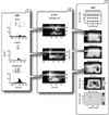
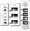
Results: Analysis of the STRF structure and integrative capacity of multisensory SC neurons revealed significant modifications in the temporal response dynamics of multisensory responses (e.g., discharge durations, peak firing rates, and mean firing rates), as well as significant changes in rates of spontaneous activation and degrees of multisensory integration.
Conclusions: These results emphasize the importance of early sensory experience in the establishment of normal multisensory processing architecture and highlight the limited plastic potential of adult multisensory circuits.
Figures


Similar articles
-
Heterogeneity in the spatial receptive field architecture of multisensory neurons of the superior colliculus and its effects on multisensory integration.Neuroscience. 2014 Jan 3;256:147-62. doi: 10.1016/j.neuroscience.2013.10.044. Epub 2013 Oct 30. Neuroscience. 2014. PMID: 24183964 Free PMC article.
-
Multisensory Plasticity in Superior Colliculus Neurons is Mediated by Association Cortex.Cereb Cortex. 2016 Mar;26(3):1130-7. doi: 10.1093/cercor/bhu295. Epub 2014 Dec 31. Cereb Cortex. 2016. PMID: 25552270 Free PMC article.
-
Visual experience is necessary for maintenance but not development of receptive fields in superior colliculus.J Neurophysiol. 2005 Sep;94(3):1962-70. doi: 10.1152/jn.00166.2005. Epub 2005 May 25. J Neurophysiol. 2005. PMID: 15917326
-
Spatial receptive field organization of multisensory neurons and its impact on multisensory interactions.Hear Res. 2009 Dec;258(1-2):47-54. doi: 10.1016/j.heares.2009.08.003. Epub 2009 Aug 19. Hear Res. 2009. PMID: 19698773 Free PMC article. Review.
-
Nonvisual influences on visual-information processing in the superior colliculus.Prog Brain Res. 2001;134:143-56. doi: 10.1016/s0079-6123(01)34011-6. Prog Brain Res. 2001. PMID: 11702540 Review.
Cited by
-
The brain can develop conflicting multisensory principles to guide behavior.Cereb Cortex. 2024 Jun 4;34(6):bhae247. doi: 10.1093/cercor/bhae247. Cereb Cortex. 2024. PMID: 38879756 Free PMC article.
-
Noise-rearing disrupts the maturation of multisensory integration.Eur J Neurosci. 2014 Feb;39(4):602-13. doi: 10.1111/ejn.12423. Epub 2013 Nov 19. Eur J Neurosci. 2014. PMID: 24251451 Free PMC article.
-
Fractality of sensations and the brain health: the theory linking neurodegenerative disorder with distortion of spatial and temporal scale-invariance and fractal complexity of the visible world.Front Aging Neurosci. 2015 Jul 15;7:135. doi: 10.3389/fnagi.2015.00135. eCollection 2015. Front Aging Neurosci. 2015. PMID: 26236232 Free PMC article.
-
Faster phonological processing and right occipito-temporal coupling in deaf adults signal poor cochlear implant outcome.Nat Commun. 2017 Mar 28;8:14872. doi: 10.1038/ncomms14872. Nat Commun. 2017. PMID: 28348400 Free PMC article.
-
Multisensory simultaneity judgment and proximity to the body.J Vis. 2016;16(3):21. doi: 10.1167/16.3.21. J Vis. 2016. PMID: 26891828 Free PMC article.
References
-
- Avillac M, Deneve S, Olivier E, Pouget A, Duhamel JR. Reference frames for representing visual and tactile locations in parietal cortex. Nat Neurosci. 2005;8(7):941–949. - PubMed
-
- Calvert GA, Spence C, Stein BE. The Handbook of Multisensory Processes. Cambridge, MA: The MIT Press; 2004.
-
- Carriere BN, Royal DW, Perrault TJ, Morrison SP, Vaughan JW, Stein BE, et al. Visual deprivation alters the development of cortical multisensory integration. J Neurophysiol. 2007;98(5):2858–2867. - PubMed
Publication types
MeSH terms
Grants and funding
LinkOut - more resources
Full Text Sources
Miscellaneous

