. 2010 Apr-Jun;4(2):95-103.
doi: 10.4161/fly.4.2.11861. Epub 2010 Apr 18.

A forward genetic screen in Drosophila melanogaster to identify mutations affecting INAD localization in photoreceptor cells

Affiliations

A forward genetic screen in Drosophila melanogaster to identify mutations affecting INAD localization in photoreceptor cells

Parthena D Sanxaridis et al. Fly (Austin). 2010 Apr-Jun.

Abstract

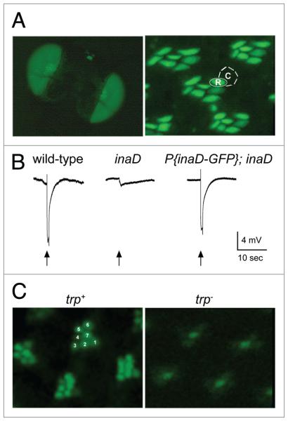
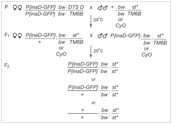
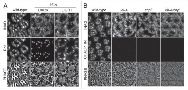
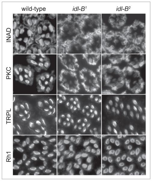
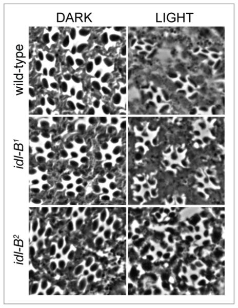
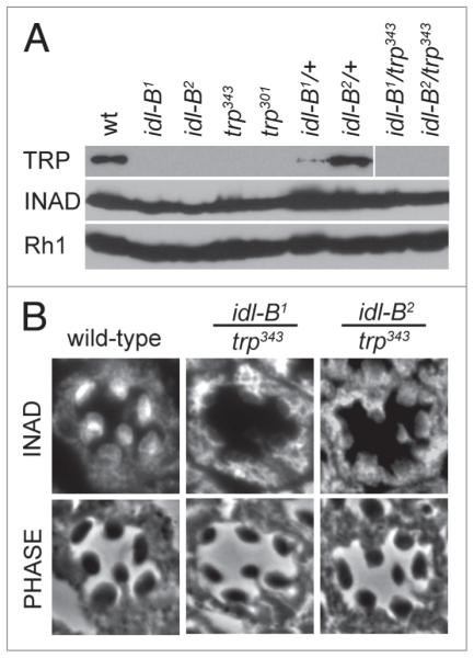
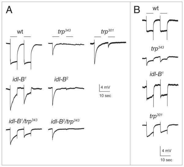
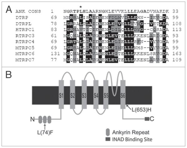
In Drosophila photoreceptors, the multivalent PDZ protein INAD interacts with multiple signaling components and localizes complexes to the rhabdomere, a subcellular compartment specialized for phototransduction. Since this localization is critical for signaling, we conducted a genetic screen of the third chromosome for mutations that result in mislocalization of an INAD-GFP fusion protein. We identified seven mutant lines that fall into two complementation groups, idl (INAD localization)-A and idl-B. We show that idl-A mutants fail to complement with chaoptic (chp) mutants. Since chaoptin is a structural component of the rhabdomere, mislocalization of INAD may be a secondary effect of the retinal degeneration in chp and idl-A mutants. Genetic complementation and DNA sequencing reveal that the two idl-B mutants represent new alleles of trp, a gene encoding the major light-activated channel. The molecular change in each allele affects a highly conserved residue in either an ankyrin domain on the N-terminus or in the S6 transmembrane domain of TRP. These changes lead to the loss of TRP protein. TRP has previously been shown to anchor INAD in the rhabdomeres, therefore the independent identification of two trp alleles validates our screen for INAD-GFP localization. One possibility is that a limited number of proteins are required for localizing INAD-signaling complexes. A similar screen of the X and second chromosomes may be required to find the remaining players involved.


Figures

Figure 1
Figure 1
INAD-GFP protein expression, localization and function in the Drosophila retina. (A) Left, INAD-GFP fusion protein is expressed in photoreceptors, as seen in whole eyes of the fly. Right, 1-μm-thick retinal cross-sections show INAD-GFP is localized to the rhabdomeres of photoreceptor cells. The rhabdomere (R) and cell body (c) of a single photoreceptor cell are approximately outlined for illustration. (B) electroretinogram (eRG) recordings from wild-type, inaD1 null, and transgenic inaD1 null flies expressing INAD-GFP (P(inaD-GFP); inaD1). Flies were exposed to a 2 second flash of light (arrow). Note that P(inaD-GFP); inaD1 flies exhibit near complete rescue of signaling. (c) Visualization of INAD-GFP in intact eyes. shown are representative ommatidia expressing INAD-GFP in inaD1 (trp+) and inaD1; trp343 (trp-) backgrounds. INAD-GFP signal is seen in all seven rhabdomeres of each ommatidia in wild-type. For reference, seven rhabdomeres are numbered in a representative ommatidium. The trp null retina displayed diffuse localization of INAD-GFP localization pattern, especially in the six outer photoreceptors.
Figure 2
Figure 2
Genetic scheme for the INAD-GFP localization screen. The genetic scheme used to generate INAD-GFP expressing mutant lines for screening. Third chromosome eMs mutagenized males from the Zuker collection (* indicates the mutation on the third chromosome) and P(inaD-GFP); bw; DTS4 D/TM6B females were crossed at 29°c for five days. The non-permissive temperature of 29°c was used to eliminate all progeny containing DTS4 D in the F1 generation. All viable F1 males and females were inbred at 25°c. DTS4 escapers (not shown in the scheme) were identified by the presence of the dominant Dichete (D) mutation. F2 homozygous mutant flies (bw; st*) were identified by the presence of white eyes and screened for INAD-GFP localization.
Figure 3
Figure 3
The idl-A mutants recovered from the screen are alleles of chaoptic. (A) shown are representative cross-sections from idl-A mutants that have been dark-raised (DARK) or light-exposed (LIGhT) for 10 days-post-eclosion, and then immunostained for INAD and Rh1. Wild-type control flies were dark-raised. A phase contrast image of each cross-section is shown to illustrate the photoreceptor structure. Wild-type retinal sections exhibited rhabdomeric localization of INAD and Rh1 in addition to robust structural integrity. Dark-raised idl-A mutant photoreceptors displayed retinal degeneration, depicted by smaller rhabdomeres, and mislocalization of INAD, but near normal localization of Rh1. With exposure to light, idl-A mutants showed a more severe loss of retinal integrity and mislocalization of both INAD and Rh1. (B) complementation analysis of idl-A and chaoptic (chp2) mutants. Retinal cross-sections from young (<2 day old), dark raised wild-type, idl-A, chp2 and idl-A/chp2 flies were immunostained for INAD and chaoptin. Wild-type photoreceptors exhibited normal retinal integrity, and rhabdomeric localization of both INAD and chaoptin. chp2, idl-A and idl-A/chp2 mutant photoreceptors all exhibited mislocalization of INAD, lack of chaoptin staining, and retinal degeneration.
Figure 4
Figure 4
INAD complexes are specifically mislocalized in idl-B mutant photoreceptor cells. shown are representative dark-raised wild-type, idl-B1 and idl-B2 retinal cross-sections (1-μm-thick) immunostained for INAD, pKc, TRpL and Rh1. Wild-type photoreceptors displayed rhabdomeric localization of INAD, pKc, TRpL and Rh1. Both idl-B alleles displayed mislocalization of INAD and pKc, but normal localization of TRpL and Rh1.
Figure 5
Figure 5
idl-B mutant photoreceptors display a light-dependant retinal degeneration. Representative retinal sections from wild-type, idl-B1 and idl-B2 flies that were dark-raised (DARK) or light-exposed (LIGHT) until 3 days-post-eclosion (3DPE ). All dark-raised flies exhibited wild-type photoreceptor structure. Light-exposure of both idl-B alleles resulted in retinal degeneration, especially depicted by smaller rhabdomeres.
Figure 6
Figure 6
idl-B mutants lack TRp protein. (A) Representative immunoblots of wild-type (wt), idl-B alleles, trp alleles, idl-B heterozygotes and trans-heterozygous idl-B/trp flies. TRp protein was absent from all homozygous trp and idl-B alleles. The two idl-B alleles failed to complement with trp as depicted by the lack of TRp protein in the trans-heterozygous idl-B/trp samples. INAD and Rh1, in contrast, were present in all fly stocks. All samples contained 10 heads from darkraised flies. (B) Representative cross-sections of single ommatidia from dark-raised wild-type, idl-B1/trp343, and idl-B2/trp343 flies immunostained for INAD. Note that INAD is mislocalized in both idl-B1/trp343 and idl-B2/trp343, indicating that both idl-B alleles failed to complement with trp for INAD localization. phase-contrast images are shown to illustrate the wild-type structure of all three genotypes analyzed.
Figure 7
Figure 7
electroretinogram recordings of idl-B mutants. (A) Representative eRG recordings from young, dark-raised wild-type and mutant flies. With white light (400–700 nm) at an intensity of 453 lux, both idl-B mutants showed a transient eRG response, similar to trp mutants. idl-B1/trp343 and idl-B2/trp343 trans-heterozygotes also displayed an eRG waveform closely resembling the trp mutants. 10 second light stimuli are depicted by a line above the eRG traces. (B) At a lower light intensity of 6.39 lux, idl-B1 responded more similar to wild-type then the trp343 null mutant. The trp301 hypomorph also responded more similar to wild-type at the lower light intensity.
Figure 8
Figure 8
idl-B mutants encode new alleles of trp. (A) Ankyrin repeat comparison. sequence alignment of ankyrin repeats (AR) from various TRpc channels and AR containing proteins against the AR consensus sequence (ANK cONs, top). The well conserved amino acids within the ANK cONs are shown in bold. Drosophila melanogaster TRp protein (DTRp; p19334); Drosophila melanogaster TRpL (DTRpL; p48994); Mus musculus TRpc5 (MTRpc5; Q9QX29); Rattus norvegicus TRpc4 (RTRpc4; O35119); Homo sapiens TRpc1 (hTRpc1; p48995); Mus musculus TRpc7 (MTRpc7; Q9WVc5); Rattus norvegicus TRpc3 (RTRpc3; Q9JM19); Mus musculus TRpc6 (MTRpc6; Q61143). The accession numbers for each protein are shown in parenthesis. NcBI BLAsT was used for protein sequence comparison and the alignment was done via clustal2W, online program. Residues identical to the consensus sequence are shaded in black, residues similar to the consensus sequence are shaded gray. The asterisk indicates the leucine at position six of the AR that is mutated in idl-B1. Many of the TRp proteins from the TRpc family were found to contain this conserved leucine within the ankyrin repeat. (B) A cartoon representation of the Drosophila TRp protein illustrating the amino acid changes resulting in each idl-B mutant. shown are the six transmembrane domains (s1-s6), three ankyrin repeats on the N-terminus and the INAD binding site at the c-terminal end of the Drosophila TRp protein. The idl-B1 mutation is an amino acid change L(74)F in the second ankyrin domain on the cytoplasmic N-terminus of TRp. The idl-B2 mutation produces an amino acid change L(653)h in the sixth transmembrane domain (s6) of TRp.

Similar articles

Cited by

References

    1. Tsunoda S, Zuker CS. The organization of INAD-signaling complexes by a multivalent PDZ domain protein in Drosophila photoreceptor cells ensures sensitivity and speed of signaling. Cell Calcium. 1999;26:165–71. - PubMed
    1. Huber A. Scaffolding proteins organize multimolecular protein complexes for sensory signal transduction. The European journal of neuroscience. 2001;14:769–76. - PubMed
    1. Ranganathan R, Ross EM. PDZ domain proteins: scaffolds for signaling complexes. Curr Biol. 1997;7:770–3. - PubMed
    1. Tsunoda S, Sierralta J, Zuker CS. Specificity in signaling pathways: assembly into multimolecular signaling complexes. Curr Opin Genet Dev. 1998;8:419–22. - PubMed
    1. Hardie RC, Raghu P. Visual transduction in Drosophila. Nature. 2001;413:186–93. - PubMed

Publication types

MeSH terms

LinkOut - more resources