Review
. 2010 May;6(5):276-87.
doi: 10.1038/nrneurol.2010.37. Epub 2010 Apr 20.

Signals to promote myelin formation and repair

Affiliations
Review

Signals to promote myelin formation and repair

Carla Taveggia et al. Nat Rev Neurol. 2010 May.

Abstract

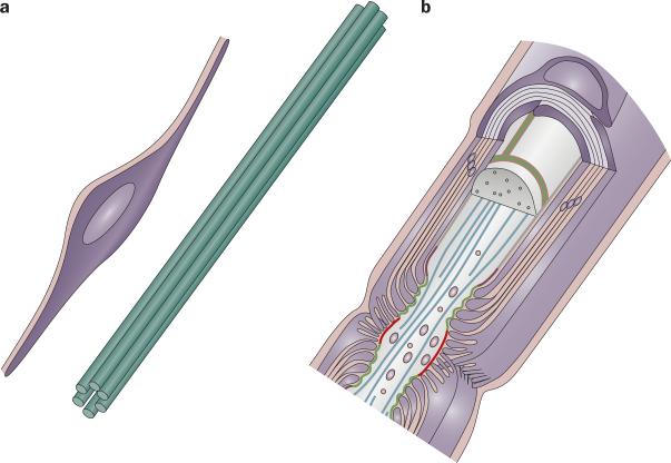
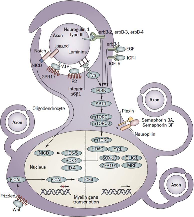
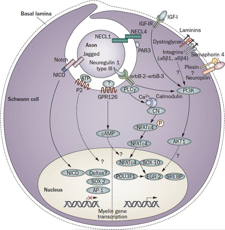
The myelin sheath wraps large axons in both the CNS and the PNS, and is a key determinant of efficient axonal function and health. Myelin is targeted in a series of diseases, notably multiple sclerosis (MS). In MS, demyelination is associated with progressive axonal damage, which determines the level of patient disability. The few treatments that are available for combating myelin damage in MS and related disorders, which largely comprise anti-inflammatory drugs, only show limited efficacy in subsets of patients. More-effective treatment of myelin disorders will probably be accomplished by early intervention with combinatorial therapies that target inflammation and other processes-for example, signaling pathways that promote remyelination. Indeed, evidence suggests that such pathways might be impaired in pathology and, hence, contribute to the failure of remyelination in such diseases. In this article, we review the molecular basis of signaling pathways that regulate myelination in the CNS and PNS, with a focus on signals that affect differentiation of myelinating glia. We also discuss factors such as extracellular molecules that act as modulators of these pathways. Finally, we consider the few preclinical and clinical trials of agents that augment this signaling.


Figures

Figure 1
Figure 1
The myelin–axon unit. The development and maintenance of the myelin–axon unit, here illustrated in the PNS, is dependent on reciprocal signaling between glia and axons. a | Schwann cells sit at the edge of a bundle of axons in prenatal nerves and provide trophic support to neurons (1). In turn, axons and other sources (for example, extracellular matrix) transmit signals that promote the survival and differentiation of glia, as well as myelination (2). b | Eventually, Schwann cells myelinate a segment of one axon. Once the myelin–axon has formed, glia transmit signals that promote axonal health (3), while signals originating from the axon and extracellular matrix promote myelin maintenance (4). Reprinted from Neuron, 40, Salzer, J. L., Polarized domains of myelinated axons, 297–318 © 2003, with permission from Elsevier.
Figure 2
Figure 2
Axon to glia signaling pathways in myelination. Schematic representation of the main signaling pathways that regulate the onset of myelination during CNS development. The figure depicts an oligodendrocyte just before wrapping. The pathways shown are not comprehensive, but focus on signaling that converges on the glial cell nucleus. Regulatory feedback loops are not displayed. For some molecules, the localization shown is putative. Note that, as oligodendrocytes must integrate signals from multiple axons, with different caliber and electrical activity, a layer of local peri-axonal regulation is likely to be superimposed on nuclear regulation of myelination. Dashed lines indicate signals with uncertain targets. Abbreviations: Nrg: Neuregulin; EGF: Epidermal Growth Factor; IGF1: Insulin-like Growth Factor 1; Sema: Semaphorin; Fyn: Fyn kinase; GPR17: G protein-coupled receptor 17; P2: Purinergic Receptors 2; NICD: Notch-1 intracellular domain: PI3K: phosphatidylinositol-3 kinase; Akt: serine/threonine-specific protein kinaseAkt/PKB; mTOR: mammalian target of rapamycin (mTOR) signaling complexes; HDAC: Histone deacetylase; YY1: YIN-YANG-1; Sox: SRY-box containing transcription factor; Zfp: zinc finger protein; MRF: Myelin gene regulatory factor; Tcf: T-cell factor 4 transcription factor; Hes: hairy and enhancer of split 5 transcription factor; Id: Inhibitor of differentiation transcription factor; β-cat: β-catenin; Wnt: Wingle wingless-related mouse mammary tumor virus integration site protein.
Figure 3
Figure 3
Axon to glia signaling in myelination. Schematic representation of the main pathways that regulate myelination during PNS development. The figure depicts a promyelinating Schwann cell. The pathways shown are not comprehensive, but focus on signaling that converges on the nucleus. Regulatory feedback loops are not displayed. For some molecules, the localization shown is putative. Dashed lines indicate signals with uncertain targets. NFkB (nuclear factor κB); Pou (Pituitary Octamer Unc-86) 3F1, also known as Tst-1/Oct6/SCIP, Egr (Early growth response) 2, also known as Krox-20; Sox ( SRY-box containing) 10 and SREBP (Sterol Regulatory Element Binding Protein) are transcription factors that activate PNS myelination, . Mutations in these genes are associated with CMT neuropathies, Waardenburg-Hirschsprung disease and central dysmyelination, . Sox (SRY-box containing) 2 and 4, Id (Inhibitor of differentiation)2, Pax3 and c-Jun-of the activator protein 1 (AP-1 complex, are inhibitory transcription factors active before myelination. Their inappropriate activation might be harmful in neuropathies. Nrg: Neuregulin; IGF1: Insulin-like Growth Factor 1; Cadm: cell adhesion molecules, also known as IGSF4, SynCAM, Necl, TSLC; PAR: Partition defective 3; GPR: G protein-coupled receptor; P2: Purinergic Receptors 2; NICD: Notch-1 intracellular domain: PI3K: phosphatidylinositol-3 kinase; PLC γ: phospholipase γ; Sema: Semaphorin; NFAT c4: Nuclear factor of activated T-cells, cytoplasmic 4.

Similar articles

Cited by

References

    1. Salzer JL, Brophy PJ, Peles E. Molecular domains of myelinated axons in the peripheral nervous system. Glia. 2008;56:1532–40. - PubMed
    1. Nave KA, Trapp BD. Axon-glial signaling and the glial support of axon function. Annu Rev Neurosci. 2008;31:535–61. - PubMed
    1. Bozzali M, Wrabetz L. Axonal signals and oligodendrocyte differentiation. Neurochem Res. 2004;29:979–88. - PubMed
    1. Simons M, Trajkovic K. Neuron-glia communication in the control of oligodendrocyte function and myelin biogenesis. J Cell Sci. 2006;119:4381–9. - PubMed
    1. Woodhoo A, Sommer L. Development of the Schwann cell lineage: from the neural crest to the myelinated nerve. Glia. 2008;56:1481–90. - PubMed

Publication types

MeSH terms

Substances