. 2010 Apr 9;5(4):e10093.
doi: 10.1371/journal.pone.0010093.

Granulocyte colony-stimulating factor activating HIF-1alpha acts synergistically with erythropoietin to promote tissue plasticity

Affiliations

Granulocyte colony-stimulating factor activating HIF-1alpha acts synergistically with erythropoietin to promote tissue plasticity

Shih-Ping Liu et al. PLoS One. .

Erratum in

  • PLoS One. 2010;5(10). doi:10.1371/annotation/433064f4-e30a-4000-8e5a-9e8d1775d820. Su, Ching-Yuan [added]; Li, Hung [added]

Abstract

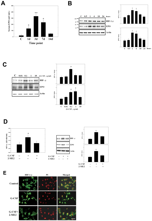
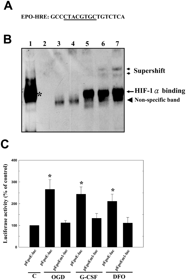
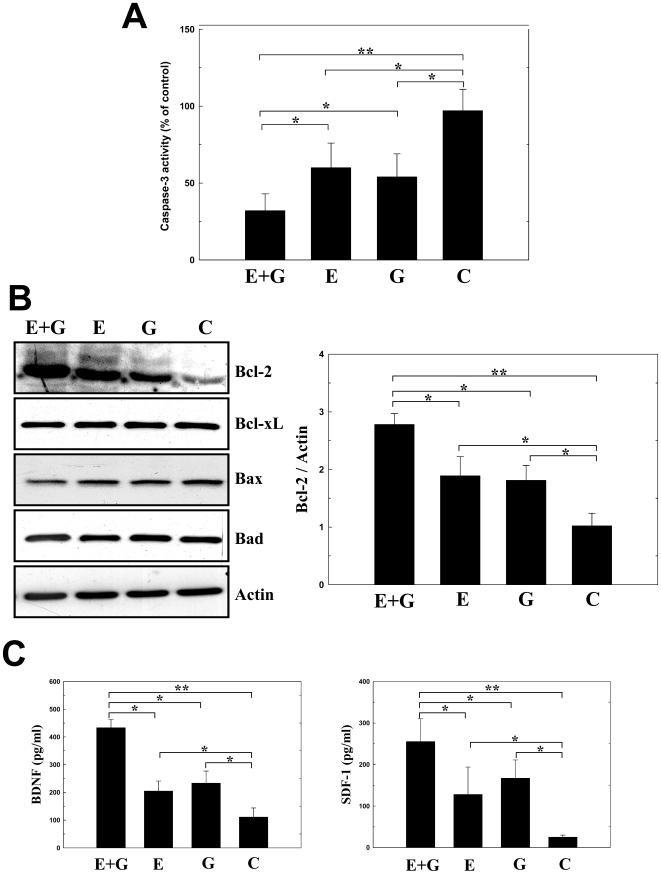
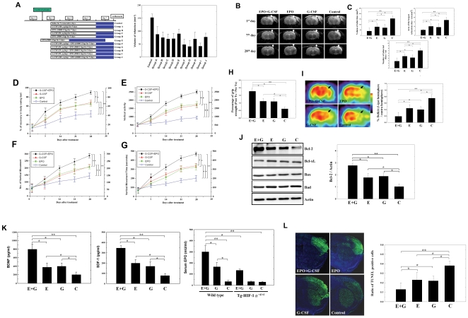
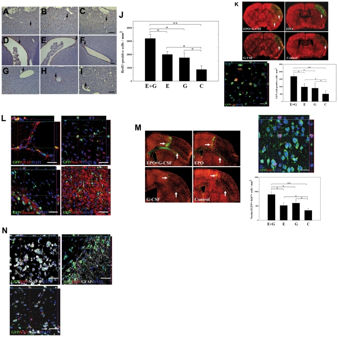
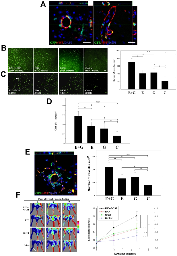
Stroke and peripheral limb ischemia are serious clinical problems with poor prognosis and limited treatment. The cytokines erythropoietin (EPO) and granulocyte-colony stimulating factor (G-CSF) have been used to induce endogenous cell repair and angiogenesis. Here, we demonstrated that the combination therapy of EPO and G-CSF exerted synergistic effects on cell survival and functional recovery from cerebral and peripheral limbs ischemia. We observed that even under normoxic conditions, G-CSF activates hypoxia-inducible factor-1alpha (HIF-1alpha), which then binds to the EPO promoter and enhances EPO expression. Serum EPO level was significantly increased by G-CSF injection, with the exception of Tg-HIF-1alpha(+f/+f) mice. The neuroplastic mechanisms exerted by EPO combined with G-CSF included enhanced expression of the antiapoptotic protein of Bcl-2, augmented neurotrophic factors synthesis, and promoted neovascularization. Further, the combination therapy significantly increased homing and differentiation of bone marrow stem cells (BMSCs) and intrinsic neural progenitor cells (INPCs) into the ischemic area. In summary, EPO in combination with G-CSF synergistically enhanced angiogenesis and tissue plasticity in ischemic animal models, leading to greater functional recovery than either agent alone.


Conflict of interest statement

Competing Interests: The authors have declared that no competing interests exist.

Figures

Figure 1
Figure 1. G-CSF increased serum EPO in human and stimulated the expression of EPO by activating HIF-1α in HUVECs.
(A) G-CSF-treatment for 5 consecutive days in human showed significant increases serum EPO level compared to control [C]. (B–C) G-CSF induced the protein expression of both HIF-1α and EPO. (D) G-CSF also enhanced the activity of HIF-1α in cell lysate. The upregulation of HIF-1α activity, and protein expression of HIF-1α and EPO stimulated by adding G-CSF returned to normal levels after addition of 2-methoxyestradiol (2-ME2). (E) G-CSF treatment induced the translocation of HIF-1α into nuclei (PI: propidium iodide, nuclear stain) or to perinuclear areas. In contrast, pretreatment with 2-ME2 inhibited the nuclear translocation of HIF-1α. Mean ± SEM, *P<0.05 and **P<0.01 vs. control. Bar  = 50 µm.
Figure 2
Figure 2. G-CSF promoted HIF-1α transcriptional activity and binding to the HRE of EPO promoter.
(A) An oligonucleotide containing the HRE of the HIF-1α-binding sequence (underlined) from the EPO gene (EPO-HRE) was used in EMSA (B), comprising positive control (black star, Lane 1), negative control (Lane 2), and DNA (DIG-labeled oligonucleotide) binding complex (Lane 5; large arrow). Binding was reduced by competition with non-labeled probe (Lane 3, 100X excess; and Lane 4, 300X excess). Nonspecific bands (arrow head) were also shown (including Land 3 and 4). For supershift assay (small arrows), polyclonal anti-HIF-1α (Lane 6) and monoclonal anti-HIF-1α (Lane 7) were used. (C) In a luciferase reporter assay, luciferase activity was higher in the G-CSF-treated cells transfected with pEpoE-luc than that with mutant pEpoEm1-luc, or the control cells. The G-CSF-stimulated luciferase activities were similar to those in oxygen glucose deprivation (OGD) and chemical hypoxia (DFO) conditions. Mean ± SEM, *P<0.05 and **P<0.01 vs. control.
Figure 3
Figure 3. EPO+G-CSF exerted an anti-apoptotic effect and enhanced neurotrophic factor synthesis in primary cortical cultures (PCCs).
(A) Under OGD, PCCs pretreated with EPO+G-CSF (E+G) resulted in less caspase-3 activity than with EPO (E) or G-CSF (G) alone, or controls (C). (B) In a Western blot analysis, EPO+G-CSF-treated PCCs expressed more Bcl-2 than EPO, G-CSF or control groups. (C) Measurement of neurotrophic factors by ELISA revealed higher levels of BDNF and SDF-1 in the EPO+G-CSF-treated PCCs than in EPO- or G-CSF-treated cells or the control. Mean ± SEM, *P<0.05 and **P<0.01 vs. control.
Figure 4
Figure 4. Administration of EPO+G-CSF to cerebral ischemic rats reduced infarct size and improved neurological function.
(A) Experimental protocols for determining the best combinations of EPO and G-CSF to reduce infarct size. The dosage, combination and injection duration in each group are indicated in the white rectangles. The blue rectangles in each group represent non-treatment day-point after cytokine injections before euthanasia. In group E, EPO 5000 U/kg treatment started 30 minutes before stroke initiation. Group C (G-CSF alone), group D (EPO alone), group I (EPO+G-CSF) and saline-control group were selected for further study. (B) Representative MRI of ischemic rat brain: the white areas (white arrows) indicate the infarct zone in the right cerebral cortex on the 1st, 7th and 28th day after cerebral infarction. (C) Three formats of cerebral infarction assessment including total infarct volume, area of largest infarct section and number of infarct sections at the 7th day after cerebral ischemia were measured in rats treated with EPO+G-CSF (E+G) than in EPO (E) or G-CSF (G)-treated rats or the control (C) group. (D-G) Body asymmetry and locomotor activities after MCA ligation were measured in rats receiving EPO+G-CSF, EPO, G-CSF, and control saline from 7 to 28 days recovery. (H) Forelimb grip strength before and after ischemia were measured in rats receiving EPO+G-CSF, EPO, G-CSF, and control saline. (I) Representative deficit (black arrow, coronal view) and semi-quantitative measurements of [18F]fluoro-2-deoxyglucose positron emission tomography (FDG-PET) images of the right cortex of EPO+G-CSF, EPO, G-CSF and control group. (J) Bcl-2, Bcl-xL, Bax, and Bad proteins expression in rats' brain were analyzed 24 hr post-cerebral ischemia following treatment with EPO+G-CSF, EPO, G-CSF or saline. (K) Neurotrophic factors BDNF and SDF-1 level in rats' brain were measured by ELISA following treatment with EPO+G-CSF EPO, G-CSF or saline. Serum EPO levels were measured by ELISA in wild-type mice (C57BL/6 mice) and Tg-HIF-1α+f/+f mice treated with G-CSF and EPO+G-CSF injection. (L) Representative TUNEL (green) and Hoechst 33342 (blue) co-staining images of cells death in right hemisphere of ischemic rat brains from EPO+G-CSF, EPO, G-CSF or control groups. Mean ± SEM, *P<0.05 and **P<0.01 vs. control. Bar  = 50 µm.
Figure 5
Figure 5. Subcutaneous administration of EPO+G-CSF enhanced the proliferation, differentiation and migration of stem cells in rats and mice.
At one week after cerebral ischemia, BrdU immunoreactive cells were detected in the ipsilateral cortex near the infarct boundary (A-C, arrows), the subventricular area (D-F, arrows), and around blood vessels (G-I, arrows). (J) Numbers of BrdU immunoreactive cells were measured in the ipsilateral hemisphere of rats' brains treated with EPO+G-CSF (E+G), EPO (E), G-CSF (G) or controls (C). (K) GFP+c-Kit+ bone marrow stem cells (BMSCs) in the peri-infarct and striatal areas (white arrows) were analyzed in transgenic GFP-chimeric mice treated with EPO+G-CSF (E+G), EPO (E), G-CSF (G) or controls. (L) In double immunofluorescent analysis (3D image), many GFP+ cells colocalized with specific markers GFAP, Neu-N, Musashi-1 and MAP-2. (M) Nestin-EGFP+ INPCs (white arrows) were stained for Ki67 in the penumbral region of nestin-EGFP mice treated with EPO+G-CSF (E+G), EPO (E), G-CSF (G) or controls. (N) Nestin-EGFP+-Ki67+ cells also co-localized with specific markers MAP-2, GFAP, and Neu-N. Mean ± SEM, *P<0.05 and **P<0.01 vs. control. Bar  = 50 µm.
Figure 6
Figure 6. EPO+G-CSF treatment induced angiogenesis.
(A) Colocalization of GFP+ and vWF+ cells in transgenic GFP-chimeric mice treated with EPO+G-CSF was analyzed in the perivascular and endothelial regions of the ischemic hemispheres. (B) Representative images of FITC-dextran perfused vessels in ischemic brain following EPO+G-CSF, EPO, G-CSF-treated and control rats. (C) Representative images and quantitative analysis of the cerebral blood vessel density after staining by CD31 immunoreactivity (white arrow) were analyzed from ischemic brain in the EPO+G-CSF, EPO, or G-CSF-treated and control rats. (D) Cerebral blood flow (CBF) was measured by laser doppler flowmetry (LDF) in the ischemic cortex treated with EPO+G-CSF, EPO, G-CSF or controls. (E) In 3D images, GFP+ cells colocalized with vascular phenotype vWF+ cells in the perivascular region of the ischemic limb muscles in hindlimb ischemic mice model. CD31-immunoreactive blood vessel density was quantified in the limb muscles treated with EPO+G-CSF, EPO, G-CSF or controls. (F) Representative images of blood perfusion in ischemic limbs were analyzed by laser doppler perfusion imaging (LDPI) analysis in mice treated with EPO+G-CSF, EPO, G-CSF or controls. Mean ± SEM, *P<0.05 and **P<0.01 vs. control. Bar  = 50 µm.

Similar articles

Cited by

References

    1. Wang L, Zhang Z, Wang Y, Zhang R, Chopp M. Treatment of stroke with erythropoietin enhances neurogenesis and angiogenesis and improves neurological function in rats. Stroke. 2004;35:1732–1737. - PubMed
    1. Signore AP, Weng Z, Hastings T, Van Laar AD, Liang Q, et al. Erythropoietin protects against 6-hydroxydopamine-induced dopaminergic cell death. J Neurochem. 2006;96:428–443. - PubMed
    1. Frampton JE, Lee CR, Faulds D. Filgrastim. A review of its pharmacological properties and therapeutic efficacy in neutropenia. Drugs. 1994;48:731–760. - PubMed
    1. Lee ST, Chu K, Jung KH, Ko SY, Kim EH, et al. Granulocyte colony-stimulating factor enhances angiogenesis after focal cerebral ischemia. Brain Res. 2005;1058:120–128. - PubMed
    1. Park HK, Chu K, Lee ST, Jung KH, Kim EH, et al. Granulocyte colony-stimulating factor induces sensorimotor recovery in intracerebral hemorrhage. Brain Res. 2005;1041:125–131. - PubMed

Publication types

MeSH terms

Substances