

Clonal analysis in mice underlines the importance of rhombomeric boundaries in cell movement restriction during hindbrain segmentation
- PMID: 20404937
- PMCID: PMC2853563
- DOI: 10.1371/journal.pone.0010112
Clonal analysis in mice underlines the importance of rhombomeric boundaries in cell movement restriction during hindbrain segmentation
Abstract
Background: Boundaries that prevent cell movement allow groups of cells to maintain their identity and follow independent developmental trajectories without the need for ongoing instructive signals from surrounding tissues. This is the case of vertebrate rhombomeric boundaries. Analysis in the developing chick hindbrain provided the first evidence that rhombomeres are units of cell lineage. The appearance of morphologically visible rhombomeres requires the segment restricted expression of a series of transcription factors, which position the boundaries and prefigure where morphological boundaries will be established. When the boundaries are established, when the cells are committed to a particular rhombomere and how they are organized within the hindbrain are important questions to our understanding of developmental regionalization.
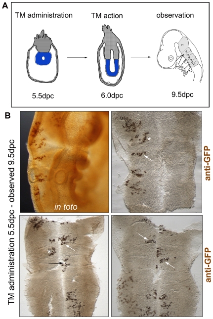
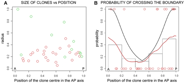
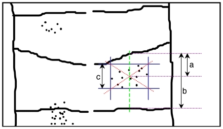
Methodology/principal findings: Sophisticated experimental tools with high-resolution analysis have allowed us to explore cell lineage restriction within the hindbrain in mouse embryos. This novel strategy is based on knock-in alleles of ubiquitous expression and allows unrestricted clonal analysis of cell lineage from the two-cell stage to the adult mouse. Combining this analysis with statistical and mathematical tools we show that there is lineage compartmentalization along the anteroposterior axis from very early stages of mouse embryonic development.
Conclusions: Our results show that the compartment border coincides with the morphological boundary in the mouse hindbrain. The restriction of the cells to cross rhombomeric boundaries seen in chick is also observed in mouse. We show that the rhombomeric boundaries themselves are involved in cell movement restriction, although an underlying pre-pattern during early embryonic development might influence the way that cell populations organize.
Conflict of interest statement
Figures
References
-
- Garcia-Bellido A, Ripoll P, Morata G. Developmental compartmentalisation of the wing disk of drosophila. Nat New Biol. 1973;245(147):251–253. - PubMed
-
- Lawrence PA. Maintenance of boundaries between developing organs in insects. Nat New Biol. 1973;242(114):31–32. - PubMed
-
- Morata G, Lawrence PA. Control of compartment development by the engrailed gene in drosophila. Nature. 1975;255(5510):614–617. - PubMed
-
- Lumsden A. Cell lineage restrictions in the chick embryo hindbrain. Philos Trans R Soc Lond B Biol Sci. 1991;331(1261):281–286. - PubMed
-
- Trainor PA, Krumlauf R. Hox genes, neural crest cells and branchial arch patterning. Curr Opin Cell Biol. 2001;13(6):698–705. - PubMed
Publication types
MeSH terms
Grants and funding
LinkOut - more resources
Full Text Sources
Molecular Biology Databases
