

The effectiveness of bone mineral density as supplementary tool for evaluation of the osteogenic potential in patients with spinal fusion
- PMID: 20404939
- PMCID: PMC2852037
- DOI: 10.4184/asj.2009.3.1.1
The effectiveness of bone mineral density as supplementary tool for evaluation of the osteogenic potential in patients with spinal fusion
Abstract
Study design: Retrospective study.
Purpose: This study was designed to determine the effectiveness of bone mineral density measurement as a supplementary tool for evaluation of osteogenic potential in patients with spinal fusion. To this end, we correlated bone mineral density (BMD) with osteogenic potential from cultured mesenchymal stem cells (MSCs).
Overview of literature: Many studies have correlated osteogenic potential of in vitro cultured MSCs with aging or osteoporosis.
Methods: We studied twenty-five individuals with harvested bone marrow from the ilium during lumbar spinal surgery. The BMD of the femoral neck was measured using dual energy X-ray absorptiometry prior to bone marrow aspiration, and the osteoporotic group was classified as those with T-scores below-2.5. After MSCs were isolated from bone marrow, in vitro induction of osteogenesis was performed. We analyzed the patient's osteogenic potential from cultured MSCs such as mineral deposition stain, bone alkaline phosphatase (ALP) activity and osteoblast-specific gene expression in RT-PCR.
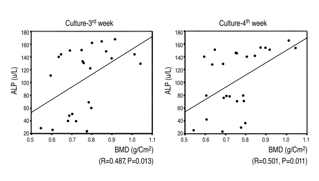
Results: On mineral staining, the osteoporotic group had a scanty matrix mineral deposition in contrast to the non-osteoporotic group. The expression of osteocalcin in the osteoporotic group was 1.5 to 3 times less than in the non-osteoporotic group. At the 3(rd) week after the induction of osteogenesis, the activity of ALP of cultured MSCs in the osteoporotic group was lower than in the control group (mean, 45+/-19 u/L, in osteoporotic group vs 136+/-7 u/L in non-osteoporotic), and there was a statistically significant and positive correlation between BMD & ALP (r=0.487, p=0.013).
Conclusions: There is a positive correlation between BMD and osteogenic potential derived from MSCs. The measurement of BMD can provide supplementary data for evaluating osteogenic potential clinically.
Keywords: Bone mineral density; Mesenchymal stem cells; Osteogenic potential.
Figures


Similar articles
-
[Osteogenic potential of bone marrow mesenchymal stem cells from ovariectomied osteoporotic rat].Sichuan Da Xue Xue Bao Yi Xue Ban. 2005 May;36(3):318-21. Sichuan Da Xue Xue Bao Yi Xue Ban. 2005. PMID: 15931856 Chinese.
-
Vertebral Bone Marrow-Derived Mesenchymal Stromal Cells from Osteoporotic and Healthy Patients Possess Similar Differentiation Properties In Vitro.Int J Mol Sci. 2020 Nov 5;21(21):8309. doi: 10.3390/ijms21218309. Int J Mol Sci. 2020. PMID: 33167522 Free PMC article.
-
Effects of Intermittent Parathyroid Hormone 1-34 Administration on Circulating Mesenchymal Stem Cells in Postmenopausal Osteoporotic Women.Med Sci Monit. 2019 Jan 8;25:259-268. doi: 10.12659/MSM.913752. Med Sci Monit. 2019. PMID: 30620727 Free PMC article.
-
Presenting a Method to Improve Bone Quality Through Stimulation of Osteoporotic Mesenchymal Stem Cells by Low-Level Laser Therapy.Photomed Laser Surg. 2017 Nov;35(11):622-628. doi: 10.1089/pho.2016.4245. Epub 2017 Jun 15. Photomed Laser Surg. 2017. PMID: 28621568 Review.
-
Relationship among Bone Mineral Density Reduction, Hearing Loss, and Balance Disorders in Osteoporotic Patients.Front Bioeng Biotechnol. 2013 Nov 25;1:17. doi: 10.3389/fbioe.2013.00017. eCollection 2013. Front Bioeng Biotechnol. 2013. PMID: 25152874 Free PMC article. Review. No abstract available.
Cited by
-
Relationship between bone mineral density and alcohol intake: A nationwide health survey analysis of postmenopausal women.PLoS One. 2017 Jun 29;12(6):e0180132. doi: 10.1371/journal.pone.0180132. eCollection 2017. PLoS One. 2017. PMID: 28662191 Free PMC article.
References
-
- Reyes M, Verfaillie CM. Characterization of multipotent adult progenitor cells, a subpopulation of mesenchymal stem cells. Ann N Y Acad Sci. 2001;938:231–233. - PubMed
-
- Bruder SP, Jaiswal N, Haynesworth SE. Growth kinetics, self-renewal, and the osteogenic potential of purified human mesenchymal stem cells during extensive subcultivation and following cryopreservation. J Cell Biochem. 1997;64:278–294. - PubMed
-
- Chomczynski P, Sacchi N. Single-step method of RNA isolation by acid guanidinium thiocyanate-phenol-chloroform extraction. Anal Biochem. 1987;162:156–159. - PubMed
-
- Gottfried ON, Dailey AT. Mesenchymal stem cell and gene therapies for spinal fusion. Neurosurgery. 2008;63:380–391. - PubMed
-
- Nakajima T, Iizuka H, Tsutsumi S, Kayakabe M, Takagishi K. Evaluation of posterolateral spinal fusion using mesenchymal stem cells: differences with or without osteogenic differentiation. Spine (Phila Pa 1976) 2007;32:2432–2436. - PubMed
LinkOut - more resources
Full Text Sources
Research Materials
Miscellaneous
