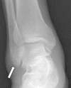
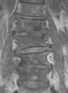
Multiple pyogenic spondylodiscitis with bilateral psoas abscesses accompanying osteomyelitis of lateral malleolus - a case report -
- PMID: 20404964
- PMCID: PMC2852090
- DOI: 10.4184/asj.2008.2.2.102
Multiple pyogenic spondylodiscitis with bilateral psoas abscesses accompanying osteomyelitis of lateral malleolus - a case report -
Abstract
A psoas abscess is a potentially life-threatening infection. Multiple pyogenic spondylodiscitis with bilateral psoas abscesses accompanying an osteomyelitis of the lateral malleolus is an extremely rare event. We present our experience with needle aspiration for the treatment of osteomyelitis of the lateral malleolus and CT-guided percutaneous catheter drainage for a psoas abscess in an elderly patient. Both infections were completely resolved without recurrence. A psoas abscess should be included in the differential diagnosis of a patient with low back pain during musculoskeletal infection. Percutaneous needle aspiration or CT-guided percutaneous catheter drainage is an effective method for treating certain musculoskeletal infections.
Keywords: CT-guided percutaneous drainage; Osteomyelitis of lateral malleolus; Spondylodiscitis with psoas abscess.
Figures


Similar articles
-
Lumbar Pyogenic Spondylodiscitis and Bilateral Psoas Abscesses Extending to the Gluteal Muscles and Intrapelvic Area Treated with CT-guided Percutaneous Drainage - A Case Report -.Asian Spine J. 2008 Jun;2(1):51-4. doi: 10.4184/asj.2008.2.1.51. Epub 2008 Jun 30. Asian Spine J. 2008. PMID: 20411143 Free PMC article.
-
CT-guided percutaneous drainage within intervertebral space for pyogenic spondylodiscitis with psoas abscess.Acta Radiol. 2012 Feb 1;53(1):76-80. doi: 10.1258/ar.2011.110418. Epub 2011 Dec 2. Acta Radiol. 2012. PMID: 22139720
-
[CT-guided percutaneous drainage in the management of psoas abscesses].Orv Hetil. 1994 Nov 20;135(47):2597-602. Orv Hetil. 1994. PMID: 7824259 Hungarian.
-
[Pyogenic psoas abscess: six cases and review of the literature].Rev Med Interne. 2006 Nov;27(11):828-35. doi: 10.1016/j.revmed.2006.07.010. Epub 2006 Aug 15. Rev Med Interne. 2006. PMID: 16959381 Review. French.
-
Pyogenic liver abscess: treatment with needle aspiration.Clin Radiol. 1997 Dec;52(12):912-6. doi: 10.1016/s0009-9260(97)80223-1. Clin Radiol. 1997. PMID: 9413964 Review.
References
-
- Gruenwald I, Abrahamson J, Cohen O. Psoas abscess: case report and review of the literature. J Urol. 1992;147:1624–1626. - PubMed
-
- Ricci MA, Rose FB, Meyer KK. Pyogenic psoas abscess: worldwide variations in etiology. World J Surg. 1986;10:834–843. - PubMed
-
- Kao PF, Tsui KH, Leu HS, Tsai MF, Tzen KY. Diagnosis and treatment of pyogenic psoas abscess in diabetic patient: usefulness of computed tomography and gallium-67 scanning. Urology. 2001;57:246–251. - PubMed
-
- Santaella RO, Fishman EK, Lipsett PA. Primary vs secondary iliopsoas abscess. Presentation, microbiology, and treatment. Arch Surg. 1995;130:1309–1313. - PubMed
-
- Dinc H, Onder C, Turhan AU, et al. Percutaneous catheter drainage of tuberculous and nontuberculous psoas abscesses. Eur J Radiol. 1996;23:130–134. - PubMed
LinkOut - more resources
Full Text Sources

