

Fluorescein and indocyanine green angiography for uveitis
- PMID: 20404985
- PMCID: PMC2855659
- DOI: 10.4103/0974-9233.58419
Fluorescein and indocyanine green angiography for uveitis
Abstract
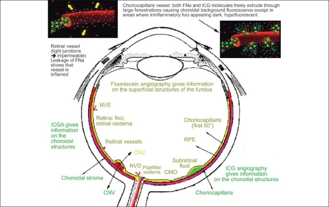
In recent years enormous progress has been achieved in investigational procedures for uveitis. Imaging is one such example with the advent of new methods such as indocyanine green angiography, ultrasound biomicroscopy and optical coherence tomography to cite only the most important. This tremendous increase in precision and accuracy in the assessment of the level and degree of inflammation and its monitoring comes in parallel with the development of extremely potent and efficacious therapies. In view of these developments, our whole attitude in the appraisal and investigation of the uveitis patient has to be adapted and correctly reoriented integrating the recent developments and this is no different for ocular angiography.
Keywords: Fluorescein; Indocyanine Green Angiography; Uveitis.
Conflict of interest statement
Figures
References
-
- De Laey JJ. Fluorescein angiography in posterior uveitis. Int Ophthalmol Clin. 1996;35:33–58. - PubMed
-
- Brancato R, Trabucchi G. Fluorescein and indocyanine green angiography in vascular chorioretinal diseases. Semin Ophthalmol. 1998;13:189–98. - PubMed
-
- Herbort CP. Posterior uveitis: New insights given by indocyanine green angiography. Eye. 1998;12:757–9. - PubMed
-
- Herbort CP, Borruat FX, de Courten C, Jaccard L. Angiographie au vert d'indocyanine dans les uvéites postérieures. Klin Monatsbl Augenheilk. 1996;208:321–6. - PubMed
-
- Walker S, Iguchi A, Jones NP. Frosted branch angeitis: A review. Eye. 2005;18:527–32. - PubMed
LinkOut - more resources
Full Text Sources
