. 2010 Apr 12;5(4):e10122.
doi: 10.1371/journal.pone.0010122.

Dysferlin interacts with tubulin and microtubules in mouse skeletal muscle

Affiliations

Dysferlin interacts with tubulin and microtubules in mouse skeletal muscle

Bilal A Azakir et al. PLoS One. .

Abstract

Dysferlin is a type II transmembrane protein implicated in surface membrane repair in muscle. Mutations in dysferlin lead to limb girdle muscular dystrophy 2B, Miyoshi Myopathy and distal anterior compartment myopathy. Dysferlin's mode of action is not well understood and only a few protein binding partners have thus far been identified. Using affinity purification followed by liquid chromatography/mass spectrometry, we identified alpha-tubulin as a novel binding partner for dysferlin. The association between dysferlin and alpha-tubulin, as well as between dysferlin and microtubules, was confirmed in vitro by glutathione S-transferase pulldown and microtubule binding assays. These interactions were confirmed in vivo by co-immunoprecipitation. Confocal microscopy revealed that dysferlin and alpha-tubulin co-localized in the perinuclear region and in vesicular structures in myoblasts, and along thin longitudinal structures reminiscent of microtubules in myotubes. We mapped dysferlin's alpha-tubulin-binding region to its C2A and C2B domains. Modulation of calcium levels did not affect dysferlin binding to alpha-tubulin, suggesting that this interaction is calcium-independent. Our studies identified a new binding partner for dysferlin and suggest a role for microtubules in dysferlin trafficking to the sarcolemma.


Conflict of interest statement

Competing Interests: The authors have declared that no competing interests exist.

Figures

Figure 1
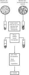
Figure 1. Schematic representation of the proteomic analysis by mass spectrometry.
The procedure includes overexpression of His-myc-dysferlin in HEK293T cells, purification on protein A-Sepharose beads coupled to anti-myc antibody, incubation of the myc-dysferlin beads with mouse skeletal muscle homogenate and the identification of the co-purified proteins using LC-MS/MS.
Figure 2
Figure 2. Alpha-tubulin is identified as a dysferlin interacting protein through affinity purification and mass spectrometry analysis.
A. Western blot to detect the expression of His-myc-dysferlin in transfected HEK293T cells (top panel), the immunoprecipitated myc-dysferlin (middle panel) and expression of endogenous dysferlin in mouse skeletal muscle homogenate (bottom panel). B. SDS-PAGE resolution of the co-purified dysferlin partner stained with silver nitrate. Left lane is the control, whereby His-myc-dysferlin was not transfected into HEK293T cells. Right lane is the immunoprecipitate with myc-dysferlin. Bands containing alpha-tubulin and previously described dysferlin binding partners are highlighted. IB: Immunoblot, IP: Immunoprecipitation, CTL: control.
Figure 3
Figure 3. Dysferlin complexes with alpha-tubulin.
A. GST, GST-TubA4A and GST-TubA1B fusion proteins immobilized onto glutathione-Sepharose 4B beads were incubated with mouse skeletal muscle homogenate. GST, GST-TubA4A or GST-TubA1B with adsorbed proteins from mouse skeletal muscle were resolved by SDS-PAGE, transferred onto a nitrocellulose membrane, and blotted with mouse monoclonal anti-dysferlin antibody. Left panel: nitrocellulose membrane stained with ponceau red, right panel: detection of immunoreactive dysferlin. B. GFP-dysferlin was overexpressed in HEK293T cells and then immunoprecipitated from cell extracts with anti-GFP antibody. As a control, protein A-Sepharose beads coated with anti-GFP antibody were incubated with extracts of non-transfected HEK293T cells. Proteins were separated on SDS-PAGE gel and were transferred onto a nitrocellulose membrane and blotted with anti-GFP or anti-alpha-tubulin antibodies. Input (right panel), immunoprecipitate (left panel). C–D. Co-immunoprecipitation of dysferlin with anti-alpha-tubulin antibody from C2C12 myotube extracts (C) or mouse skeletal muscle homogenate (D). Input (right panel), immunoprecipitate (left panel). As a control (CTL), protein A-Sepharose beads were incubated with myotube extracts in the absence of anti-alpha-tubulin antibody.
Figure 4
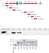
Figure 4. Alpha-tubulin interacts with dysferlin through the C2A and C2B domains.
A. Schematic illustration of full-length wild type dysferlin and the various GST-C2 domain constructs used. B. C2C12 myoblast extract was incubated with GST alone or with the various GST-C2 domains precoupled to glutathione-Sepharose 4B beads. The bound proteins were separated on SDS-PAGE followed by Western blot analysis using anti-alpha-tubulin antibody. SM: standard material. Lower panel: nitrocellulose membrane of GST-dysferlin C2 domains with adsorbed proteins from the cell extract stained with ponceau red.
Figure 5
Figure 5. Alpha-tubulin interacts with dysferlin in a calcium-independent manner.
A. Upper panel: Myoblast cell extracts were incubated with GST alone or the various GST-dysferlin C2 domain fusion proteins precoupled to glutathione-Sepharose 4B beads in the absence (−) or presence (+) of 1 mM calcium. The bound proteins were separated on SDS-PAGE followed by Western blot analysis using anti-alpha-tubulin antibody. Lower panel: nitrocellulose membrane of GST-dysferlin C2 domains with adsorbed proteins from the cell extract stained with ponceau red. B. Co-immunoprecipitation of alpha-tubulin and dysferlin with anti-alpha-tubulin antibody from mouse skeletal muscle homogenate in the presence of increasing calcium concentrations. Proteins were separated and detected with anti-alpha-tubulin and anti-dysferlin antibodies. As a control (CTL), protein A-Sepharose beads were incubated with muscle homogenate in the absence of anti-alpha-tubulin antibody.
Figure 6
Figure 6. Dysferlin binds to microtubules.
His-myc-dysferlin purified on Ni-NTA beads was incubated with polymerized microtubules. Reactions were resolved by SDS-PAGE stained with SimplyBlue SafeStain. Arrows point to His-myc-dysferlin, tubulin, BSA, and to MAP1&MAP2 of the microtubule-associated protein fraction (MAPF), which includes MAP2A, MAP2B, MAP1 and tau. S: Soluble phase, P: Pellet. Lane 1: Microtubules alone, lane 2: Microtubules incubated with MAPF, lane 3: Microtubules incubated with BSA, lane 4: MAPF alone, lane 5: BSA alone, lane 6: Purified His-myc-dysferlin alone, lane 7: Microtubules incubated with purified His-myc-dysferlin.
Figure 7
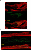
Figure 7. Alpha-tubulin co-localizes with dysferlin.
GFP-dysferlin was expressed in C2C12 myoblasts and myotubes. The localization of GFP-dysferlin was compared to that of endogenous alpha-tubulin stained with anti-alpha-tubulin antibody by confocal microscopy. The co-localization of dysferlin with alpha-tubulin is revealed in the merged image. Arrowheads point to areas of co-localization. Scale bar represents 10 µm.

Similar articles

Cited by

References

    1. Bashir R, Britton S, Strachan T, Keers S, Vafiadaki E, et al. A gene related to Caenorhabditis elegans spermatogenesis factor fer-1 is mutated in limb-girdle muscular dystrophy type 2B. Nat Genet. 1998;20:37–42. - PubMed
    1. Liu J, Aoki M, Illa I, Wu C, Fardeau M, et al. Dysferlin, a novel skeletal muscle gene, is mutated in Miyoshi myopathy and limb girdle muscular dystrophy. Nat Genet. 1998;20:31–36. - PubMed
    1. Illa I, Serrano-Munuera C, Gallardo E, Lasa A, Rojas-Garcia R, et al. Distal anterior compartment myopathy: a dysferlin mutation causing a new muscular dystrophy phenotype. Ann Neurol. 2001;49:130–134. - PubMed
    1. Therrien C, Dodig D, Karpati G, Sinnreich M. Mutation impact on dysferlin inferred from database analysis and computer-based structural predictions. Journal of the Neurological Sciences. 2006;250:71–78. - PubMed
    1. Vandre DD, Ackerman WEt, Kniss DA, Tewari AK, Mori M, et al. Dysferlin is expressed in human placenta but does not associate with caveolin. Biol Reprod. 2007;77:533–542. - PubMed

Publication types