. 2010 Apr 12;5(4):e10125.
doi: 10.1371/journal.pone.0010125.

A vertebrate-specific Chp-PAK-PIX pathway maintains E-cadherin at adherens junctions during zebrafish epiboly

Affiliations

A vertebrate-specific Chp-PAK-PIX pathway maintains E-cadherin at adherens junctions during zebrafish epiboly

Hwee Goon Tay et al. PLoS One. .

Abstract

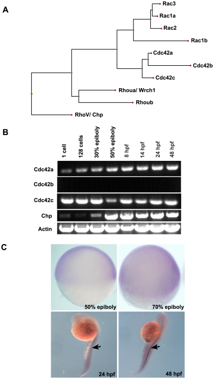
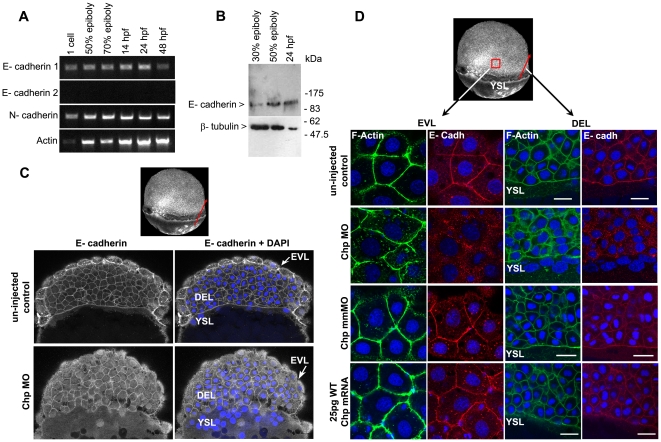
Background: In early vertebrate development, embryonic tissues modulate cell adhesiveness and acto-myosin contractility to correctly orchestrate the complex processes of gastrulation. E-cadherin (E-cadh) is the earliest expressed cadherin and is needed in the mesendodermal progenitors for efficient migration. Regulatory mechanisms involving directed E-cadh trafficking have been invoked downstream of Wnt11/5 signaling. This non-canonical Wnt pathway regulates RhoA-ROK/DAAM1 to control the acto-myosin network. However, in this context nothing is known of the intracellular signals that participate in the correct localization of E-cadh, other than a need for Rab5c signaling.

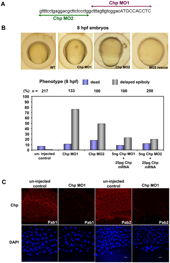
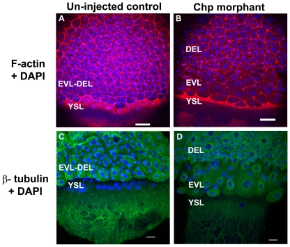
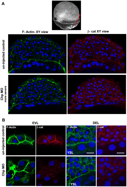
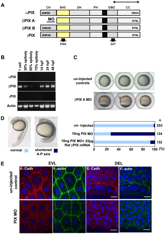
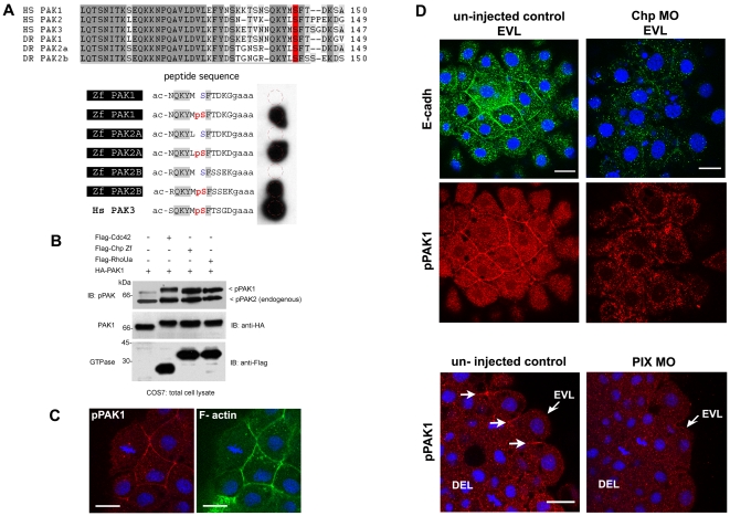
Methodology/principal findings: By studying loss of Chp induced by morpholino-oligonucleotide injection in zebrafish, we find that the vertebrate atypical Rho-GTPase Chp is essential for the proper disposition of cells in the early embryo. The underlying defect is not leading edge F-actin assembly (prominent in the cells of the envelope layer), but rather the failure to localize E-cadh and beta-catenin at the adherens junctions. Loss of Chp results in delayed epiboly that can be rescued by mRNA co-injection, and phenocopies zebrafish E-cadh mutants. This new signaling pathway involves activation of an effector kinase PAK, and involvement of the adaptor PAK-interacting exchange factor PIX. Loss of signaling by any of the three components results in similar underlying defects, which is most prominent in the epithelial-like envelope layer.

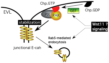
Conclusions/significance: Our current study uncovers a developmental pathway involving Chp/PAK/PIX signaling, which helps co-ordinate E-cadh disposition to promote proper cell adhesiveness, and coordinate movements of the three major cell layers in epiboly. Our data shows that without Chp signaling, E-cadh shifts to intracellular vesicles rather than the adhesive contacts needed for directed cell movement. These events may mirror the requirement for PAK2 signaling essential for the proper formation of the blood-brain barrier.


Conflict of interest statement

Competing Interests: The authors have declared that no competing interests exist.

Figures

Figure 1
Figure 1. Protein sequence analysis and spatio-temporal expression of Chp.
(A) The relationship between Chp, Cdc42 and Rac in zebrafish (Danio rerio). Protein sequences were aligned using ClustalW (DNAStar) and the dendrogram generated by Phylip. Accession numbers for cDNA sequences of Cdc42 and Rac family are: Rac1a, AY865568. Rac1b, XP_001332092.1. Rac2, AY865569. Rac3, AY865570. Cdc42a, AY865566. Cdc42b, XM_678979. Cdc42c, AY865567. Rhoua, AY865564. Rhoub, NM_001017784, Chp/ RhoV, NM_001012250. (B) RT-PCR products showing mRNA transcript profile of zebrafish Chp at different developmental stages as indicated. Both Cdc42a and Cdc42c were detected throughout the stages tested. Chp transcripts appear at epiboly. (C) Whole mount in situ hybridization (WISH) using dioxygenin (DIG) anti-sense Chp RNA probes indicates all cell types express the transcript at 50% and 80% epiboly (lateral views): Chp mRNA is enriched in notochord (indicated by arrows) at 24 hpf and 48 hpf.
Figure 2
Figure 2. Chp function is required for epibolic movements.
(A) Positions and sequence of Chp morpholino-oligoncleotides used in the study. The Chp MO1 and MO2 (corresponding to the reverse strand) are represented in purple and green respectively; lowercase sequence is 5′ UTR region. (B) Phenotypic examination of Chp morphants at 8 hpf revealed delayed closure of the yolk plug when compared with the un-injected controls. Rescue of the phenotype was performed by co-injecting Chp MO 1/2 with WT Chp (Zf) mRNA. Co-injection of 25 pg Chp mRNA with 5 ng Chp MO1/2 efficiently reduced the number of yolk plug defects at 8 hpf. Phenotypic analysis shows significant rescue in the delay of yolk plug closure at 8 hpf. (C) Immuno-histochemical staining of un-injected controls and Chp morphants at 60% epiboly using Chp antibody raised and purified from two animals (Pab1 and Pab2). Confocal images show endogenous Chp is strongly depleted Chp morphants. Decreased protein levels were observed in EVL, DEL and YSL layers with Chp MO. Embryos were counter-stained with DAPI. Scale bars = 20 µm.
Figure 3
Figure 3. The actin cytoskeleton and microtubule networks in Chp morphants.
Low resolution confocal images (10× objective) comparing (A) un-injected control and (B) Chp morphant with respect to F-actin organization at the vegetal margins of EVL and DEL, and at the external yolk syncytial layer (YSL). The deep cells marked by DAPI stained nuclei, fail to properly migrate in Chp morphants (B and D). As a result the cell margin is thinner and the F-actin ring of the EVL and YSL appears more compact. Confocal images at higher magnification (40×) comparing (C) un-injected control and (D) Chp morphant stained with β-tubulin. Microtubule organization in Chp morphant was largely normal. Scale bars represent 40 µm in panels A-B and 20 µm in panels C-D.
Figure 4
Figure 4. E-cadh is not maintained at the AJs in the absence of Chp.
(A) RT-PCR products of zebrafish E-cadh1, E-cadh2 and N-cadherin (N-cadh) at the stages shown. E-cadh1 transcript was present throughout all stages however we did not detect expression of E-cadh2 (although it is represented 3 times in the EST database). N-cadh transcripts accumulate more significantly at 50% epiboly. Primers encoding the cytoplasmic domain of E-cadh1 NM_131820, E-cadh2 XM_690906 and N-cadh AF 418565 were used. (B) Immunoblot analysis of endogenous E-cadh at the stages indicated. The E-cadh antibody (BD Biosciences) detects a single E-cadh band (approx. 120kDa). (C) Low resolution image of phalloidin stained embryo of 60% epiboly, marked with red arrow representing the ‘y’ component in xy cross-section of the embryo (EVL→ DEL→ YSL) where the confocal image is taken. A middle sagittal plane of the embryo at 60% epiboly is derived from the Z stack. E-cadh was no longer maintained at the AJs in the EVL and was found predominately in cytoplasm. (D) Schematic diagram (3D view) of 60% epiboly. Red box indicates the lateral area of EVL and red arrow indicates the ‘y vector’ of the xy confocal slice. Confocal images (40× mag.) showing E-cadh co-localized with F-actin at the AJs between adjacent cells in the EVL and DEL. These observations were similar with the controls that were injected with 25 pg WT Chp mRNA alone and Chp mmMO. The images represent a stack of 10 images (each 0.5 µm); E-cadh staining was rarely detected at AJs in Chp morphant but intracellular signal was not diminished, suggesting E-cadh is mis-localized without Chp signaling. Identical loss of E-cadh was observed (data not shown) with a Mab that recognized the extracellular domain of E-cadh (ECM Biosciences; CM1681). Scale bars represent 20 µm.
Figure 5
Figure 5. Localization of β-cat at the AJs requires Chp signaling.
(A) A middle sagittal plane of the embryo at 60% epiboly immunostained with β-cat is also derived from the Z stack. β-cat was no longer maintained at the AJs in the EVL and was found predominately in cytoplasm. Scale bars represent 20 µm. (B) Immunostaining of β-cat comparing the EVL and DEL of un– injected control and Chp morphant. Levels of anti-β-cat staining at AJs were severely reduced in Chp morphants. The delocalized β-cat protein was detected within intracellular structures, but no nuclear enrichment was observed relative to controls.
Figure 6
Figure 6. PIX is required to localize E-cadh at the AJs.
(A) Schematic of zebrafish PIX isoforms designated αPIX, βPIX-A, βPIX-B and the newly described γPIX. Arrows indicate the positions of the oligonucleotide primers used for RT-PCR. The position of the PIX-MO at the 5′ UTR of βPIX-A is indicated: this transcript encodes the smaller PIX isoform which is equivalent to the ubiquitous mammalian βPIX. SH3 domain in yellow and GBD binds to PAK and GIT respectively. (B) Transcript profile showing RT-PCR products for PIX at the developmental stages indicated. Primers cover essentially the same region of the PIX ORFs and therefore do not discriminate between the alternate spliced forms at the 5′ terminus. (C) PIX morphant embryos exhibit epibolic delay compared to un-injected controls at 8 hpf. (D) The typical phenotype of embryos depleted of βPIX-A at 24 hpf, exhibit shortened AP axes suggesting gastrulation defects. Phenotypic analysis showing significant rescue at 24 hpf after co-injection with 25 pg of rat βPIX mRNA. (E) Reduced cell junctional E-cadh signals in the EVL and DEL after PIX knock-down. The level of cortical F-actin (phalloidin) is similar to controls but the junctional network is more irregular. Intracellular E-cadh puncta suggest PIX functions downstream of Chp to maintain E-cadh at cell adhesions. Scale bars = 20 µm.
Figure 7
Figure 7. PAK lies downstream of Chp signaling and active kinase co-localizes with E-cadh.
(A) Characterization of the phospho-PAK (pPAK) antibody. Alignment of auto-inhibitory domain (AID) of human (Hs) and zebrafish (Zf) PAKs: HsPak1, NP_001122092.1; HsPak2, NP_002568.2; HsPak3, NP_002569.1; ZfPak1, NP_958485.1; ZfPak2a, NP_001002717; and ZfPak2b, NP_001020627.1. Dark and light grey shaded sequences represent identical and conserved amino acids; the serine residue highlighted in red is the phosphorylated site recognized by the rabbit anti-pPAK144 antibody. Further characterization of this new antibody will be presented elsewhere; it is ∼10 times more sensitive than the anti-PAK1 pSer199 described previously in zebrafish . Anti-pS144PAK1 recognizes all zebrafish PAK isoforms as assessed by synthetic phospho-peptides representing zebrafish PAK sequences. The peptides were synthesized in situ on cellulose (Jerinini) with a 3 amino-acid linker at the C-terminal end, and N-terminally amidated. The filter was blocked with BSA, and probed with anti-pPAK and HRP-anti-rabbit IgG antibodies both at 1∶2000 (30 min each, with 3×10 min washes). Human PAK3 (pS139) is shown as a positive control. (B) Constitutively active Chp (G38V) can activate PAK1. Active Cdc42(G12V), Chp(G38V) and RhoUa(Q104L) were cloned in the mammalian expression vector pXJ-Flag (with CMV promoter) and co-expressed with HA-tagged PAK1 in COS-7 cells. The activation of PAK1 is indicated by an upshift in the PAK1 band and by PAK1 phosphorylation on Ser144. (C) Activated pPAK1 was detected at the centrosome of mitotic cell (as previously reported in mammalian cell culture) and can be found at the cell junctions of envelope cells (EVL), but not on the junctions of deeper cells. (D) E-cadh colocalizes with pPAK1 at the AJs of the EVL but not in the cytoplasmic puncta. Both were reduced at the AJs of the EVL in Chp morphants. Junctional pPAK signal is reduced in the envelope cells in PIX morphants. Typical stainings of control and PIX morphant embryos. Both images represent a stack of 3 confocal images, collected under the same laser and gain settings, and at equivalent positions in the embryo. White arrows represent junctional pPAK1. Scale bars represent 20 µm.
Figure 8
Figure 8. Model of the Chp-PAK-PIX mediated pathway uncovered in this study, and its possible location with reference to other components controlling E-cadh localization at the EVL of epiboly.
Wnt11 has been shown to control E-cadh via a Rab5 sensitive pathway .

Similar articles

Cited by

References

    1. Babb SG, Marrs JA. E-cadherin regulates cell movements and tissue formation in early zebrafish embryos. Dev Dyn. 2004;230:263–277. - PubMed
    1. Montero JA, Carvalho L, Wilsch-Brauninger M, Kilian B, Mustafa C, et al. Shield formation at the onset of zebrafish gastrulation. Development. 2005;132:1187–1198. - PubMed
    1. Ulrich F, Krieg M, Schotz EM, Link V, Castanon I, et al. Wnt11 functions in gastrulation by controlling cell cohesion through Rab5c and E-cadherin. Dev Cell. 2005;9:555–564. - PubMed
    1. Shimizu T, Yabe T, Muraoka O, Yonemura S, Aramaki S, et al. E-cadherin is required for gastrulation cell movements in zebrafish. Mech Dev. 2005;122:747–763. - PubMed
    1. Kane DA, McFarland KN, Warga RM. Mutations in half baked/E-cadherin block cell behaviors that are necessary for teleost epiboly. Development. 2005;132:1105–1116. - PubMed

Publication types

Substances