

Prevalence of talon cusps in Jordanian permanent teeth: a radiographic study
- PMID: 20406435
- PMCID: PMC2861631
- DOI: 10.1186/1472-6831-10-6
Prevalence of talon cusps in Jordanian permanent teeth: a radiographic study
Abstract
Background: The aim of the study is to investigate the prevalence of talon cusps in a sample of Jordanians dental patients and their distribution among different types of teeth.
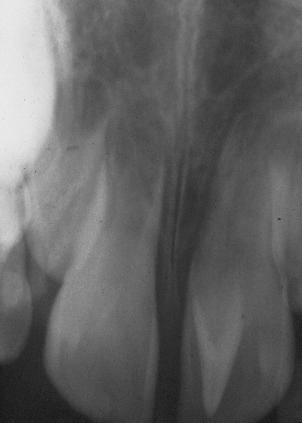
Methods: The data were collected from radiographic examination of 3,024 periapical films showing 9,377 teeth from a random sample of 1,660 patients. A tooth was considered having talon cusp if there was a V-shape radiopaque structure superimposed the tooth structure.
Results: Talon cusps were detected in 52 teeth (tooth prevalence = 0.55%). Maxillary canines were the most commonly affected teeth (46% of cases), followed by maxillary lateral incisor teeth (39% of cases) and maxillary central incisors teeth (15% of cases). Teeth with talon cusps were found in 40 subjects (person prevalence = 2.4%). Bilateral talon cusps were seen in 12 patients.
Conclusions: Attention should be paid to the presence of talon cusp and the treatment problems associated with it.
Figures
References
-
- Peker I, Alkurt MT. Talon cusp: a case series. Gen Dent. 2009;57:524–527. - PubMed
-
- Hattab FN, Yassin OM, al-Nimri KS. Talon cusp in permanent dentition associated with other dental anomalies: review of literature and reports of seven cases. ASDC J Dent Child. 1996;63:368–376. - PubMed
-
- Chawla HS, Tewari A, Gopalakrishnan NS. Talon cusp--a prevalence study. J Indian Soc Pedod Prev Dent. 1993;1:28–34. - PubMed
MeSH terms
LinkOut - more resources
Full Text Sources
