. 2010 Apr 20:5:23.
doi: 10.1186/1745-6150-5-23.

Tumor morphological evolution: directed migration and gain and loss of the self-metastatic phenotype

Affiliations

Tumor morphological evolution: directed migration and gain and loss of the self-metastatic phenotype

Heiko Enderling et al. Biol Direct. .

Abstract

Background: Aside from the stepwise genetic alterations known to underlie cancer cell creation, the microenvironment is known to profoundly influence subsequent tumor development, morphology and metastasis. Invasive cluster formation has been assumed to be dependent on directed migration and a heterogeneous environment--a conclusion derived from complex models of tumor-environment interaction. At the same time, these models have not included the prospect, now supported by a preponderance of evidence, that only a minority of cancer cells may have stem cell capacity. This proves to weigh heavily on the microenvironmental requirements for the display of characteristic tumor growth phenotypes. We show using agent-based modeling that some defining features of tumor growth ascribed to directed migration might also be realized under random migration, and discuss broader implications for cause-and-effect determination in general.

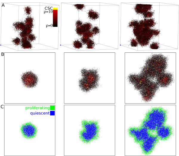
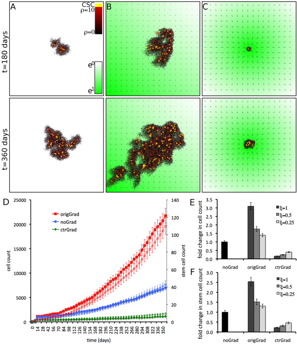
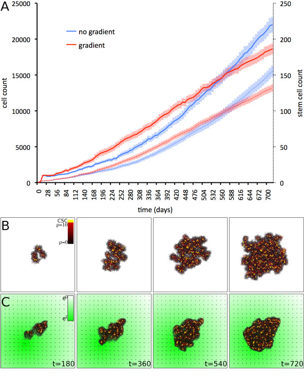
Results: Considering only the properties of random migration in tumors composed of stem cells and committed cells, we are able to recapitulate a characteristic clustering feature of invasive tumor growth, a property we attribute to "self-metastatic" growth. When the additional influence of directed migrations under chemotactic environments are considered, we find that tumor growth and invasive morphology are supported while the tumor is distant from the source, but are progressively discouraged as the tumor converges about that source.

Conclusions: We show that invasive clustering can derive from basic kinetic assumptions often neglected in more complex models. While higher-order mechanisms, e.g. directed migration upon chemotactic stimuli, may result in clustering growth morphologies, exclusive attributions of this phenotype to this or other structured microenvironments would be inappropriate, in light of our finding these features are observable in a homogeneous environment. Furthermore, directed migration will result in loss of the invasive phenotype as the tumor approaches the attractor source.

Reviewers: This article was reviewed by Mark Little and Glen Webb.


Figures

Figure 1
Figure 1
Random motility and self-metastatic growth. A) Visualization of self-metastatic growth at three different time points in three-dimensional space. Individual tumor clusters are driven by cancer stem cells (yellow), and each cluster features a radial proliferation capacity fall off (red to black). B) Self-metastatic growth in two-dimensional space. C) Distribution of proliferating and quiescent cells in the tumor populations shown in B. Other parameters are μ = 15, ps = 1.
Figure 2
Figure 2
Taxis dependent tumor size and morphology. Tumor growth simulated for t = 360 days from a single cancer stem cell located the center of the computational domain (A, C) or in the center of the top right quarter P(width*3/4, height*3/4) (B). The strength of the attractor is shown in green, and green arrows show the direction and strength of local gradients. Yellow cells are cancer stem cells, and red-black shows the cellular remaining proliferation capacity ρ. A) Homogeneous domain, random motility only. B) Attractor at the origin of the computational domain P(0,0), ξ = 1. C) Attractor at center of the computational domain P(width/2, height/2), ξ = 1. D) Change in cell number over time for n = 25 simulations each of tumor growth in domains without gradients (blue circles), center gradient (green diamonds) and origin gradient (red squares). Respective numbers of stem cells are shown as thin plots (open markers). Shown are averages and standard error. E) Fold change in cell count after t = 360 days in different domains for various chemotactic response strengths ξ. F) Fold change in stem cell count after t = 360 days in different domains for various chemotactic response strengths ξ. Other parameters are ρ = 10, μ = 5, ps = 10.
Figure 3
Figure 3
Biphasic influence of directed migration. Comparison of simulations of tumor growth without environmental gradients (random migration only; blue plots) to tumor growth with directed migration up a gradient to a nearby attractor (red plots). A) During directed migration towards an attractor source the tumor grows faster due to continuous stem cell liberation. Once the tumor has reached the attractor source intra-tumoral competition for space due to inbound directed migration slows down tumor growth. The biphasic influence is observed in total cell count (solid lines) and stem cell count (dotted lines). B) Representative simulation of self-metastatic tumor growth without environmental gradients. C) Representative simulation of tumor growth initiated at P(width*1/3, height*1/3) with directed migration towards an attractor source at P(width*2/3, height*2/3). The strength of the attractor is shown in green, and green arrows show the direction and strength of local gradients. An initial growth phase towards the attractor is followed by growth around the attractor. Other parameters are ρ = 10, μ = 5, ps = 10.

References

    1. Folkman J. Tumor angiogenesis: therapeutic implications. N Engl J Med. 1971;285(21):1182–6. - PubMed
    1. Almog N, Ma L, Raychowdhury R, Schwager C, Erber R, Short S, Hlatky L, Vajkoczy P, Huber PE, Folkman J, Abdollahi A. Transcriptional Switch of Dormant Tumors to Fast-Growing Angiogenic Phenotype. Cancer Res. 2009;69(3):836–44. doi: 10.1158/0008-5472.CAN-08-2590. - DOI - PubMed
    1. Hanahan D, Weinberg R. The hallmarks of cancer. Cell. 2000;100(1):57–70. doi: 10.1016/S0092-8674(00)81683-9. - DOI - PubMed
    1. Al-Hajj M, Wicha MS, Benito-Hernandez A, Morrison SJ, Clarke MF. Prospective identification of tumorigenic breast cancer cells. Proc Natl Acad Sci USA. 2003;100(7):3983–8. doi: 10.1073/pnas.0530291100. - DOI - PMC - PubMed
    1. Fioriti D, Mischitelli M, Di Monaco F, Di Silverio F, Petrangeli E, Russo G, Giordano A, Pietropaolo V. Cancer stem cells in prostate adenocarcinoma: a target for new anticancer strategies. J Cell Physiol. 2008;216(3):571–5. doi: 10.1002/jcp.21493. - DOI - PubMed

Publication types

LinkOut - more resources