. 2010 Jun 25;285(26):19935-46.
doi: 10.1074/jbc.M109.079574. Epub 2010 Apr 20.

Probing the specificity of binding to the major nuclear localization sequence-binding site of importin-alpha using oriented peptide library screening

Affiliations

Probing the specificity of binding to the major nuclear localization sequence-binding site of importin-alpha using oriented peptide library screening

Sundy N Y Yang et al. J Biol Chem. .

Abstract

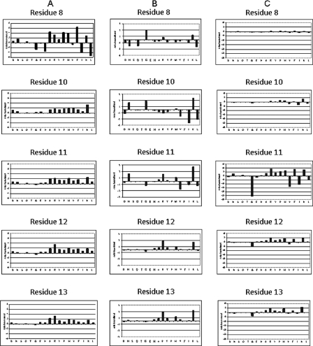
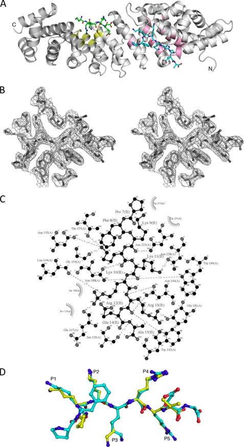
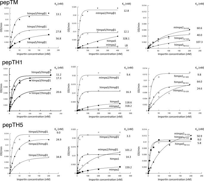
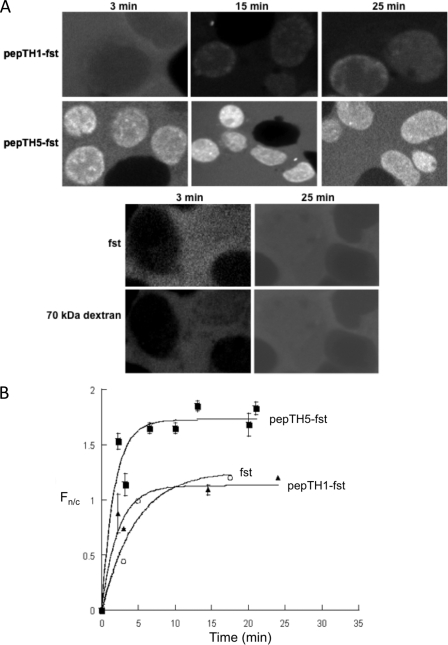
Importin-alpha is the nuclear import receptor that recognizes the classic monopartite and bipartite nuclear localization sequences (cNLSs), which contain one or two clusters of basic amino acids, respectively. Different importin-alpha paralogs in a single organism are specific for distinct repertoires of cargos. Structural studies revealed that monopartite cNLSs and the C-terminal basic clusters of the bipartite cNLSs bind to the same site on importin-alpha, termed the major cNLS-binding site. We used an oriented peptide library approach with five degenerate positions to probe the specificity of the major cNLS-binding site in importin-alpha. We identified the sequences KKKRR, KKKRK, and KKRKK as the optimal sequences for binding to this site for mouse importin-alpha2, human importin-alpha1, and human importin-alpha5, respectively. The crystal structure of mouse importin-alpha2 with its optimal peptide confirmed the expected binding mode resembling the binding of simian virus 40 large tumor-antigen cNLS. Binding assays confirmed that the peptides containing these sequences bound to the corresponding proteins with low nanomolar affinities. Nuclear import assays showed that the sequences acted as functional cNLSs, with specificity for particular importin-alphas. This is the first time that structural information has been linked to an oriented peptide library screening approach for importin-alpha; the results will contribute to understanding of the sequence determinants of cNLSs, and may help identify as yet unidentified cNLSs in novel proteins.


Figures

FIGURE 1.
FIGURE 1.
Oriented peptide library screening. A, specificity of binding to mImpα270–529. A degenerate peptide library (sequence GSEFESPXKXXXXEA, where X indicates any amino acid except Cys and Trp), was used to screen binding to GST-mImpα270–529 bound to beads. Each panel shows the relative abundance of each of the 18 amino acids in a given cycle of sequencing of eluted peptides, compared with control GST beads in the same cycle. B, same as in A, but for hImpα167–503. C, same as in A, but for hImpα560–511.
FIGURE 2.
FIGURE 2.
Crystal structure of pepTM-mImpα270–529 complex. A, overall structure of the pepTM·mImpα270–529 complex. mImpα270–529 is shown as a ribbon diagram (pink and yellow colors correspond to the residues defining the major and minor cNLS-binding sites, respectively). The superhelical axis of the repetitive part of the molecule is approximately horizontal. The two cNLS peptides are shown in a stick representation; the peptide bound to the major site is colored cyan, and the peptide bound to the minor site is colored green. B, stereoview of the electron density (drawn with the program PyMOL) in the region of the pepTM peptide bound to the major binding site of mImpα70–529. All peptide residues were omitted from the model, and simulated annealing was run with the starting temperature of 1000 K. The electron density map was calculated with coefficients 3|Fobs| − 2|Fcalc| and data between 40 and 2.3 Å resolution, and contoured at 1.2 standard deviations. Superimposed is the refined model of the peptide. C, schematic diagram of the interactions between the pepTM peptide and the major binding site of mImpα70–529. Polar contacts are shown with dashed lines, and hydrophobic contacts are indicated by arcs with radiating spokes. The cNLS peptide residues are labeled with “R.” Carbon, nitrogen, and oxygen atoms are shown in black, white, and gray, respectively; prepared with the program LIGPLOT. D, superposition of the peptide in the major cNLS binding site in the pepTM- (cyan) and T-ag- (yellow) mImpα70–529 (PDB ID 1EJL) complexes. The Ca atoms of mImpα70–529 in the two complex structures were used in the superposition.
FIGURE 3.
FIGURE 3.
Specificity of Impα binding to optimal cNLSs. Binding of Imps to the peptides as determined using an ELISA-based binding assay. Microtiter plates were coated with the peptide and incubated with increasing concentrations of Imps as indicated. Data were fitted to the function B(x) = Bmax(1 − ekt), where x is the concentration of Imp and B is the level of Imp bound. Results shown are from a single typical experiment, performed in triplicate. See Table 4 for pooled data. Top row, pepTM; middle row, pepTH1; and bottom row, pepTH5.
FIGURE 4.
FIGURE 4.
Impα-binding peptides are functional as NLSs in in vitro nuclear import assays. Nuclear import was reconstituted in perforated hepatoma tissue culture cells in the presence of an ATP-regenerating system containing GTP, GDP, and GDP-loaded Ran. Nuclear accumulation of pepTH1- and pepTH5-coupled streptavidin (fst) in the presence of hImpα5·hImpβ1 was followed over time by using a confocal laser scanning microscope. A, confocal laser scanning microscope images are shown for the indicated times at room temperature in the presence of RanGDP. B, results from image analysis are shown, where each data point represents at least five separate measurements for each of Fn, Fc, and background fluorescence. Data are fitted to the function Fn/c = Fn/cmax (1 − ekt), where Fn/cmax is the maximal level of nuclear accumulation, k is the rate constant, and t is time in minutes. Filled squares, pepTH5-fst; open circles, pepTH1-fst; and filled triangles, fst. Pooled data are presented in Table 5.
FIGURE 5.
FIGURE 5.
Correlation of nuclear import efficiency with importin binding affinity. Reciprocal plot of selected data from Tables 4 and 5 for the binding affinity (nanomolar) (y axis) and initial nuclear import rate (Fn/cinit) in minutes (x axis); the initial import rate value was determined after subtraction of the import rate for fst alone (no NLS). The line represents linear regression (r = 0.8925).

Similar articles

Cited by

References

    1. Aitchison J. D., Wozniak R. W. (2007) Nature 450, 621–622 - PubMed
    1. Alber F., Dokudovskaya S., Veenhoff L. M., Zhang W., Kipper J., Devos D., Suprapto A., Karni-Schmidt O., Williams R., Chait B. T., Sali A., Rout M. P. (2007) Nature 450, 695–701 - PubMed
    1. Kobe B., Kajava A. V. (2000) Trends Biochem. Sci. 25, 509–515 - PubMed
    1. Lange A., Mills R. E., Lange C. J., Stewart M., Devine S. E., Corbett A. H. (2007) J. Biol. Chem. 282, 5101–5105 - PMC - PubMed
    1. Cingolani G., Petosa C., Weis K., Müller C. W. (1999) Nature 399, 221–229 - PubMed

Publication types

MeSH terms

Associated data

LinkOut - more resources