. 2010 Jun 18;285(25):19184-92.
doi: 10.1074/jbc.M110.115147. Epub 2010 Apr 20.

Apobec-1 complementation factor modulates liver regeneration by post-transcriptional regulation of interleukin-6 mRNA stability

Affiliations

Apobec-1 complementation factor modulates liver regeneration by post-transcriptional regulation of interleukin-6 mRNA stability

Valerie Blanc et al. J Biol Chem. .

Abstract

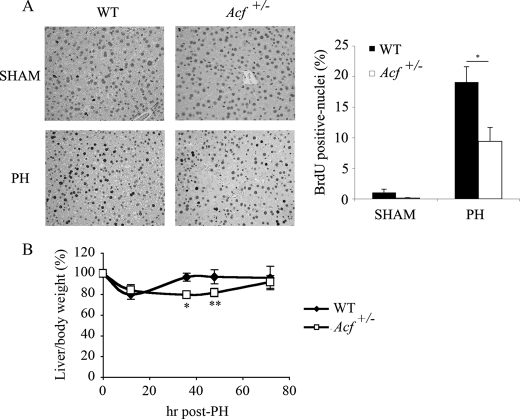
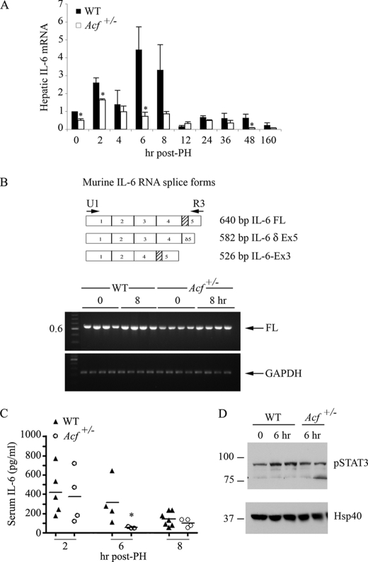
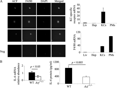
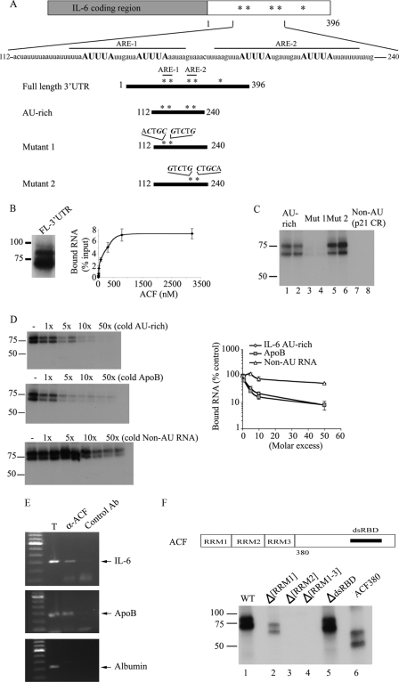
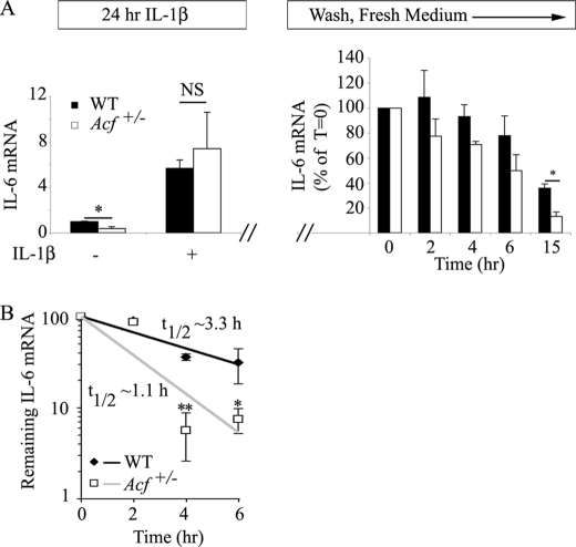
Apobec-1 complementation factor (ACF) is the RNA binding subunit of a core complex that mediates C to U RNA editing of apolipoprotein B (apoB) mRNA. Targeted deletion of the murine Acf gene is early embryonic lethal and Acf(-/-) blastocysts fail to implant and proliferate, suggesting that ACF plays a key role in cell growth and differentiation. Here we demonstrate that heterozygous Acf(+/-) mice exhibit decreased proliferation and impaired liver mass restitution following partial hepatectomy (PH). To pursue the mechanism of impaired liver regeneration we examined activation of interleukin-6 (IL-6) a key cytokine required for induction of hepatocyte proliferation following PH. Peak induction of hepatic IL-6 mRNA abundance post PH was attenuated >80% in heterozygous Acf(+/-) mice, along with decreased serum IL-6 levels. IL-6 secretion from isolated Kupffer cells (KC) was 2-fold greater in wild-type compared with heterozygous Acf(+/-) mice. Recombinant ACF bound an AU-rich region in the IL-6 3'-untranslated region with high affinity and IL-6 mRNA half-life was significantly shorter in KC isolated from Acf(+/-) mice compared with wild-type controls. These findings suggest that ACF regulates liver regeneration following PH at least in part by controlling the stability of IL-6 mRNA. The results further suggest a new RNA target and an unanticipated physiological function for ACF beyond apoB RNA editing.


Figures

FIGURE 1.
FIGURE 1.
Impaired liver regeneration in Acf+/− mice after partial hepatectomy. A, left panel, sections from wild type and Acf+/− mice livers harvested 48 h after partial hepatectomy and stained for BrdUrd incorporation. Right panel, percentage of hepatocytes labeled with BrdUrd 48 h following partial hepatectomy in wild type and Acf+/− mice (6–8 animals per genotype; *, p < 0.05). B, reduced liver to body weight ratio in Acf+/− mice following PH. Wild type and Acf+/− animals were weighed and sacrificed 12, 36, 48, and 72 h after PH and liver weights recorded. Data show the liver to body weight ratio, normalized to the starting liver to body weight ratio values (5.8% liver weight/body weight for WT animals versus 5.4% for Acf+/− mice). Data are from four to five animals per genotype, per time point; *, p < 0.05; **, p < 0.01.
FIGURE 2.
FIGURE 2.
Altered IL-6 gene expression profile during liver regeneration in Acf+/− mice. A, wild-type and Acf+/− mice were sacrificed at the indicated times following PH and RNA extracted. Expression profile of IL-6 mRNA (individual samples) was analyzed by quantitative reverse transcription-PCR. Data were standardized to the expression of 18 S mRNA (n = 3–8 animals per genotype and time point; *, p < 0.05). B, top panel, scheme of alternative splicing of murine IL-6 RNA. Shaded box indicates the deleted region resulting from alternative splicing of exon 5. Lower panel, IL-6 reverse transcription-PCR profile from WT and Acf+/− mice at 0 and 8 h following liver resection. Specific primers U1 and R3 (see “Materials and Methods” and panel) were used to co-amplify the three IL-6 spliced forms. PCR products were separated on a 1.8% agarose gel and visualized by ethidium bromide under UV light. cDNAs from 4 animals per genotype and per condition were analyzed. Only the full-length (FL) form of IL-6 RNA was detected. GAPDH (glyceraldehyde-3-phosphate dehydrogenase) mRNA was amplified and used as control. C, reduced serum IL-6 in Acf+/− mice. Serum was collected at the indicated times following PH and serum IL-6 was analyzed by enzyme-linked immunosorbent assay. D, STAT3 activation after partial hepatectomy. Phosphorylated STAT3 protein was analyzed by Western blot in liver extracts from WT and Acf+/− that were sacrificed 6 h after liver resection. Hsp40 was used as loading control.
FIGURE 3.
FIGURE 3.
ACF expression in Kupffer cells modulates IL-6 production. A, left panel, purified Kupffer cells were seeded on coverslips overnight and co-stained with rabbit anti-ACF IgG, and mouse F4/80. 4′,6-Diamidino-2-phenylindole staining indicates the nucleus. This is a representative of two independent assays. Here are shown three separate fields. Right panels, Kupffer cell specific-receptor mRNA and F4/80 mRNA expression in liver (Liv), hepatocytes (Hep), Kupffer cells (KCs), and mouse peritoneal macrophages (PMs) were analyzed by Q-PCR. Data were standardized to 18 S RNA. Data represent mean ± S.E. (n = 3–5 RNA isolations per cell type or tissue). B, Kupffer cells, isolated from WT and Acf+/− mice, were grown overnight at 37 °C. Medium and RNA were collected for IL-6 RNA and protein analyses. IL-6 RNA was analyzed by Q-PCR. 18 S RNA was used to standardize the data. Data represent mean ± S.E. (n = 4–5 Kupffer cell isolations per genotype). KC medium was used in an enzyme-linked immunosorbent assay for IL-6 quantitation, the data represent mean ± S.E. (n = 3 Kupffer cell isolations per genotype).
FIGURE 4.
FIGURE 4.
Characteristics of 3′-UTR of murine IL-6 mRNA and binding activity of ACF. A, the 3′-UTR of murine IL-6 contains 5 copies of the consensus sequence AUUUA, indicated as asterisks. The primary sequence of a 128-nt motif surrounding the AUUUA motifs is shown. Numbering begins with the first nucleotide of the 3′-UTR of IL-6 mRNA. ARE-1 extends from nucleotides 126 to 151. ARE-2 extends from nucleotides 160 to 191. Several IL-6 3′-UTR RNA templates were constructed. The 128-nt AU-rich template contains the proximal 4 AUUUA motifs. Mutants 1 and 2 contain point mutations, respectively, in the first two and last two AUUUA motifs. All the point mutations substituted the A and U nucleotides for C and G (“Materials and Methods”). B, left panel, in vitro transcribed radiolabeled IL-6 full-length (FL) 3′-UTR was incubated with 25 ng of recombinant ACF and subjected to UV cross-linking. The cross-linked products were analyzed by SDS-10% PAGE. Right panel, determination of ACF binding affinity to IL-6 full-length 3′-UTR. Increasing amounts of recombinant ACF were incubated with radiolabeled IL-6 full-length 3′-UTR probe (see “Materials and Methods”). The bound RNA was quantitated after binding to nitrocellulose filters. The apparent Kd was calculated as the concentration of ACF required to retain 50% of the maximum bound RNA. Data represent the mean ± S.E. from 3 separate assays in which each point represents triplicate determinations. C, recombinant ACF was incubated for 15 min with in vitro transcribed AU-rich, mutant 1, mutant 2, or a non-AU template (p21 coding region) of identical size. After UV cross-linking, the samples were analyzed on SDS-10% PAGE. Mutation of the two proximal AUUUA motifs abrogates ACF interaction with IL-6 AU-rich RNA (lanes 3 and 4), whereas mutation of the distal two AUUUA motifs do not affect ACF binding to the RNA probe (lanes 5 and 6). This is representative of at least 3 separate experiments. D, left panel, increasing molar excess of unlabeled (cold) IL-6 AU-rich RNA (top) or unlabeled (cold) apoB RNA (middle) were added to the radiolabeled IL-6 AU-rich RNA probe in the reaction mixture and are shown to compete out ACF binding to the IL-6 AU-rich template with similar efficiency (2–3-fold molar excess). Bottom left panel, increasing excess molar concentration of unlabeled non-AU RNA was added to labeled IL-6 AU-rich RNA and is shown to compete out ACF binding with ∼50-fold molar excess. Right panel, quantitative representation of UV cross-link binding competition from the left panels. The data represent mean ± S.E. (n = 3–5 separate experiments per condition). E, in vivo CLIP reveals ACF-IL-6 interaction. After in vivo cross-linking, LPS-stimulated macrophage (RAW 264.3) cell extracts were incubated with either unrelated rabbit IgG (control Ab) or anti-ACF antibody. The coimmunoprecipitated RNAs were extracted from the immune complexes and used as template for reverse transcription-PCR using primers specific for IL-6. The PCR products were separated on a 1% agarose gel. Lane 1 shows IL-6 mRNA in the crude cellular extract before immunoprecipitation. The position of the IL-6 PCR product is indicated by the arrow on the right. Molecular weights (kb) are shown on the left. Similarly, ACF is shown to bind apoB RNA, its canonical RNA template (middle panel), in a CLIP assay from mouse liver but fails to interact with a non-AU rich RNA (albumin) (lower panel). This is representative of two independent assays. F, determination of ACF functional domains required for binding to IL-6 mRNA. Top panel, schematic representation of ACF functional domains (6). The three RRM and the putative double-stranded RNA binding domain (dsRBD) are shown. Bottom panel, individual recombinant ACF mutants (10 ng) were incubated with radiolabeled IL-6 AU-rich template, cross-linked, and analyzed by SDS-10% PAGE. ACF interaction with IL-6 mRNA is dependent on the presence of RRM2 and RRM3 as shown by the lack of cross-link of Δ[RRM2] and Δ[RRM1–3] mutants (lanes 3 and 4).
FIGURE 5.
FIGURE 5.
ACF stabilizes IL-6 mRNA. A, IL-6 mRNA steady state in MEFs. WT and Acf+/− MEFs were incubated in the presence of 1 ng/ml of IL-1β for 24 h. Total RNA was isolated from two independent preparations of MEFs. Baseline IL-6 mRNA was significantly (*, p = 0.018) lower in MEFs from Acf+/− mice, but both genotypes responded to IL-1β administration with a 5.7–7.4-fold increase in IL-6 mRNA in WT and Acf+/− mice, respectively. In a separate series of studies, following 1 ng/ml of IL-1β incubation for 24 h, MEFs were subjected to 2 washes with PBS, subsequently maintained in normal growth medium, and total RNA isolated at the indicated time points. IL-6 mRNA levels were determined by quantitative reverse transcription-PCR and normalized to glyceraldehyde-3-phosphate dehydrogenase. Data represent mean ± S.E. from 4 independent assays per genotype (*, p = 0.033). B, KCs were isolated from WT and Acf+/− mice and plated at 4 × 10⁵ cells per well in a 12-well plate. Following an overnight culture at 37 °C, medium was changed and supplemented with LPS (final concentration 500 ng/ml) for 1 h. Conditioned medium was collected, fresh medium containing actinomycin D (Act D) (10 μg/ml) was added, and RNA was harvested at the indicated time points. IL-6 mRNA levels were determined by quantitative reverse transcription-PCR and normalized to glyceraldehyde-3-phosphate dehydrogenase mRNA. The t½ of IL-6 mRNA was 1.1 ± 0.14 h in Acf+/− KCs versus 3.3 ± 0.44 h in WT KCs. Data represent the mean ± S.E. from three independent experiments per genotype. Each experiment was performed with KCs isolated from three to four mice. *, p = 0.033; **, p = 0.0069.

References

    1. Glisovic T., Bachorik J. L., Yong J., Dreyfuss G. (2008) FEBS Lett. 582, 1977–1986 - PMC - PubMed
    1. Blanc V., Davidson N. O. (2003) J. Biol. Chem. 278, 1395–1398 - PubMed
    1. Teng B., Burant C. F., Davidson N. O. (1993) Science 260, 1816–1819 - PubMed
    1. Mehta A., Kinter M. T., Sherman N. E., Driscoll D. M. (2000) Mol. Cell. Biol. 20, 1846–1854 - PMC - PubMed
    1. Lellek H., Kirsten R., Diehl I., Apostel F., Buck F., Greeve J. (2000) J. Biol. Chem. 275, 19848–19856 - PubMed

Publication types

LinkOut - more resources