. 2010 May 15;123(Pt 10):1716-23.
doi: 10.1242/jcs.062661. Epub 2010 Apr 20.

MUC1 regulates nuclear localization and function of the epidermal growth factor receptor

Affiliations

MUC1 regulates nuclear localization and function of the epidermal growth factor receptor

Benjamin G Bitler et al. J Cell Sci. .

Abstract

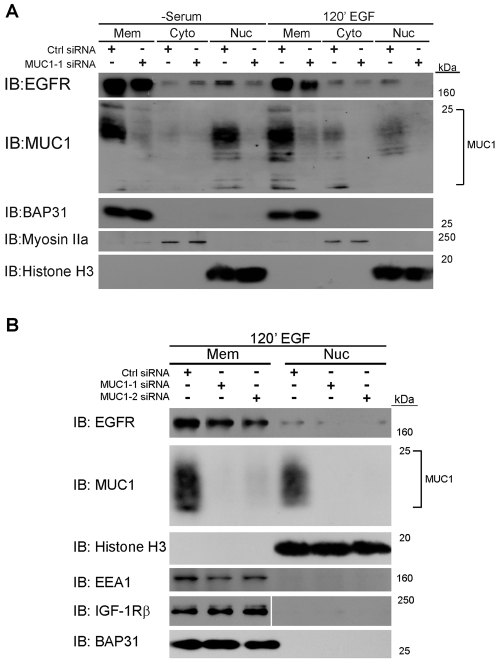
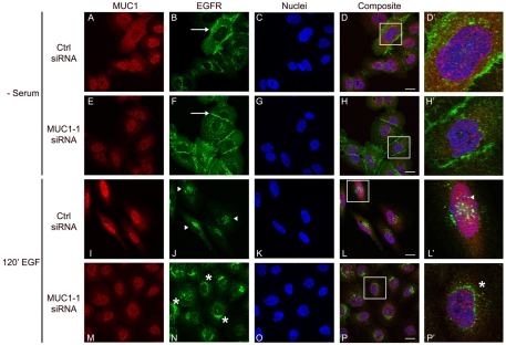
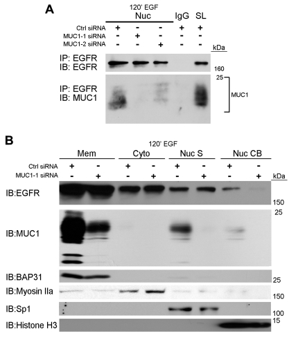
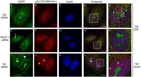
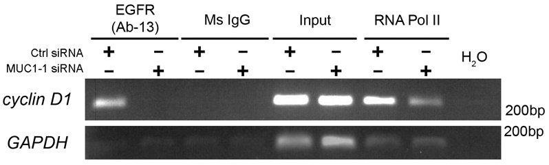
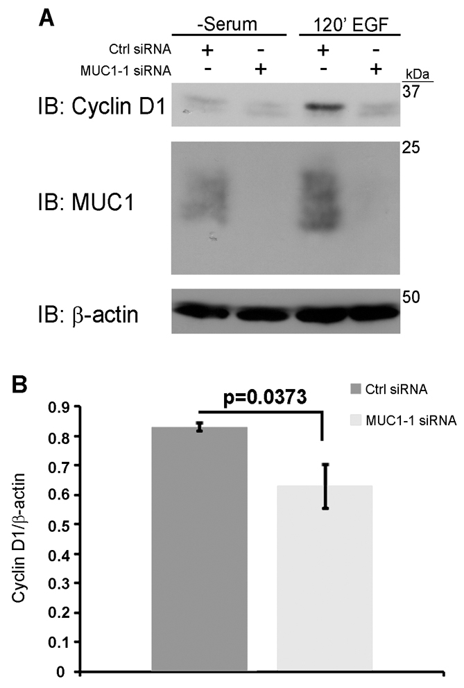
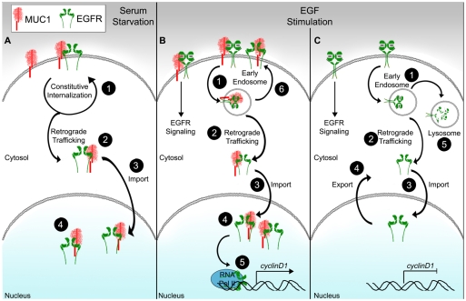
Alteration of protein trafficking and localization is associated with several diseases, including cystic fibrosis, breast cancer, colorectal cancer, leukemia and diabetes. Specifically, aberrant nuclear localization of the epidermal growth factor receptor (EGFR), a receptor tyrosine kinase, is a poor prognostic indicator in several epithelial carcinomas. It is now appreciated that in addition to signaling from the plasma membrane, EGFR also trafficks to the nucleus, and can directly bind the promoter regions of genes encoding cyclin D1 (CCND1) and B-Myb (MYBL2). We have previously established that loss of MUC1 in an EGFR-dependent transgenic mouse model of breast cancer correlates with the loss of cyclin D1 expression. Here, we provide evidence for a novel regulatory function of MUC1 in the trafficking and nuclear activity of EGFR. We found that MUC1 and EGFR interact in the nucleus of breast cancer cells, which promotes the accumulation of chromatin-bound EGFR. Additionally, the presence of MUC1 results in significant colocalization of EGFR and phosphorylated RNA polymerase II, indicating that MUC1 influences the association of EGFR with transcriptionally active promoter regions. Importantly, we found that the loss of MUC1 expression resulted in a decrease in the interaction between EGFR and the CCND1 promoter, which translated to a significant decrease in cyclin D1 protein expression. This data offers insights into a novel regulatory mechanism of EGFR nuclear function and could have important implications for evaluating nuclear localization in cancer.


Figures

Fig. 1.
Fig. 1.
MUC1 promotes nuclear localization of EGFR. (A) MDA-MB-468 breast cancer cells or (B) MCF10a immortalized breast epithelial cells treated for 3 days with control (Ctrl) or MUC1 (MUC1-1 or MUC1-2) siRNA, were either serum starved (−serum) for 18 hours or serum starved and then treated with EGF (10 ng/ml, 120 minutes). Cells were fractionated and protein (6 μg) was separated by SDS-PAGE. After transfer to PVDF, proteins were immunoblotted (IB) for EGFR (Santa Cruz, 1005) and MUC1 (Neomarkers, CT2). Fraction purity and loading were determined by immunoblotting for BAP31(Affinity Bio-Reagents, MA3-002; ER Membrane, Mem), IGF-1Rβ (Santa Cruz, SC-713; plasma membrane), EEA1 (Santa Cruz, SC-33585; Endosomes), myosin IIa (Santa Cruz, D16; cytosol, Cyto), and histone H3 (Santa Cruz, C-16; Nucleus, Nuc). White lines through blots indicate same samples and exposure but were non-contiguous. Molecular masses (kDa) are indicated on the right. Note that the cytoplasmic domain of MUC1 runs as several species of 14-28 kDa, because of different glycosylation states (Schroeder et al., 2001).
Fig. 2.
Fig. 2.
MUC1 promotes nuclear EGFR accumulation. Immortalized breast epithelial cells (MCF10A) were treated with MUC1-1 (E-H′, M-P′) or control (Ctrl) siRNA (A-D′, I-L′) and stimulated with EGF (20 ng/ml) for 120 minutes. (I-P′) Cells were fixed, permeabilized and used for EGFR immunostaining (Neomarkers, Ab-1) (green, B,F,J,N) and MUC1 (Neomarkers, CT2) (red, A,E,I,M). (A-H′) Under serum-starvation conditions (− serum) EGFR localized to the plasma membrane (white arrows), nucleus and cytoplasm. (I-L) In the presence of EGF and MUC1, EGFR is localized within the nucleus (white arrowheads, L′). (M-P′) EGF treatment in the absence of MUC1 resulted in EGFR localization to perinuclear regions (asterisk, P′). DAPI, blue nuclei. Scale bars: 20 μm.
Fig. 3.
Fig. 3.
MUC1-EGFR interaction in the nucleus promotes EGFR-chromatin interaction. (A) MCF10A immortalized breast epithelial cells were treated with control or MUC1-1 siRNA. Cells were serum starved and treated with EGF (10 ng/ml, 120 minutes at 37°C). The nuclear protein fraction (400 μg) was used for immunoprecipitation (IP) with antibodies against EGFR (Neomarkers, Ab-13) and mouse IgG, and the precipitated protein was separated by SDS-PAGE and immunoblotted (IB) for EGFR (Santa Cruz, 1005) and MUC1 (Neomarker, CT2). SL, straight lysate (non-IP protein, 12 μg). Fraction purity was confirmed by immunoblotting for histone H3 (Santa Cruz, C-16) and myosin IIa (Santa Cruz, D16). Note that straight lysates for this experiment are shown in Fig. 1B. (B) MDA-MB-468 breast cancer cells were fractionated into membrane (Mem), cytosolic (Cyto), nuclear-soluble (Nuc S) and chromatin-bound (Nuc CB) fractions and separated (15 μg) by SDS-PAGE and immunoblotted (IB) for EGFR (Santa Cruz, 1005) and MUC1 (Neomarker, CT2). Fraction purity and loading was determined by immunoblotting for BAP31 (American Bio-Reagents, MA3-002), myosin IIa (Santa Cruz, D16), Sp1 (Santa Cruz, 1C6) and histone H3 (Santa Cruz, C-16). Molecular masses (kDa) are indicated on the side.
Fig. 4.
Fig. 4.
EGFR colocalizes with phosphorylated RNA polymerase II. MCF10A cells were treated with MUC1-1 (E-H′) or control (Ctrl) siRNA (A-D′, I-L′) and stimulated with 20 ng/ml EGF for 120 minutes. Following treatment, the cells were fixed, permeabilized and incubated with antibodies against EGFR (Santa Cruz, 1005) (green, A,E,I) and phosphorylated serine-2 RNA polymerase II (pS2-CTD-RNAPolII) (Abcam, H5) (red, B,F,J). Cells were treated with DAPI to visualize nuclei (C,G,K) and composite colocalization of all three images are shown in D,H and L. (A-D) Colocalization (yellow) is seen at the arrowheads (D′). Cytoplasmic EGFR staining is shown by arrow in E. (I-L) Cells were also treated with insulin (50 ng/ml) to illustrate membrane EGFR localization (arrowhead, I) and pS2-CTD-RNA Pol II without EGF treatment (asterisks, L′). DAPI, blue nuclei. Scale bars: 10 μm.
Fig. 5.
Fig. 5.
EGFR interaction with the CCND1 promoter is MUC1 dependent. MCF10A cells were transfected with either MUC1-1 or control (Ctrl) siRNA, serum starved, treated with 10 ng/ml EGF, and incubated for 120 minutes. Protein-DNA complexes were collected and chromatin immunoprecipitations were performed against EGFR (Neomarker, AB-13), mouse IgG and RNA Pol II (Millipore, clone CTD4H8). DNA was isolated from the immunoprecipitations and used for PCR against the ATRS region of the CCND1 promoter and the GAPDH promoter.
Fig. 6.
Fig. 6.
MUC1 promotes cyclin D1 expression. (A) MCF10A cells were treated with control (Ctrl) or MUC1-1 siRNA and stimulated with 20 ng/ml EGF for 120 minutes. Cytosolic protein was isolated, analyzed by SDS-PAGE and immunoblotted for cyclin D1 (Cell Signaling, 2922), MUC1 (Neomarker, CT2) and β-actin (Sigma, AC-15). Molecular masses (kDa) are indicated on the right. (B) Relative Cyclin D1 expression levels were determined by densitometry and two-tailed Student's t-test was performed based on three experiments.
Fig. 7.
Fig. 7.
The role of MUC1 in EGFR nuclear trafficking and function. (A) Absence of EGF. (1) Constitutive internalization of MUC1 to maintain glycosylation also results in EGFR being endocytosed. (2) EGFR and MUC1 both undergo retrograde trafficking through the ER and are released into the cytosol by the Sec61 translocon. (3) Both proteins are able to interact with importin-β1, which mediates entry in the nucleus. (4) Interaction of MUC1 and EGFR in the nucleus promotes nuclear EGFR accumulation. (B) EGF stimulation in the presence of MUC1. (1) Following EGF treatment, EGFR is rapidly endocytosed in a clathrin-coated vesicle. (2) EGFR and MUC1 undergo retrograde trafficking. (3,4) Both proteins are imported into the nucleus and form a complex. (5) EGFR colocalizes with pRNA Pol II and associates with the CCND1 promoter to activate transcription. (6) Expression of MUC1 promotes the recycling of EGFR back to the plasma membrane. (C) EGF stimulation in the absence of MUC1. (1) EGF treatment results in EGFR endocytosis. EGFR is either retrograde trafficked (2) or is degraded in the lysosome (5). (3) Cytosolic EGFR is imported into the nucleus and the absence of MUC1 inhibits EGFR nuclear accumulation. (4) EGFR is then exported out of the nucleus.

Similar articles

Cited by

References

    1. Aggarwal P., Lessie M. D., Lin D. I., Pontano L., Gladden A. B., Nuskey B., Goradia A., Wasik M. A., Klein-Szanto A. J., Rustgi A. K., et al. (2007). Nuclear accumulation of cyclin D1 during S phase inhibits Cul4-dependent Cdt1 proteolysis and triggers p53-dependent DNA rereplication. Genes Dev. 21, 2908-2922 - PMC - PubMed
    1. Ahmad R., Raina D., Joshi M. D., Kawano T., Ren J., Kharbanda S., Kufe D. (2009). MUC1-C oncoprotein functions as a direct activator of the nuclear factor-{kappa}B p65 transcription factor. Cancer Res. 69, 7013-7021 - PMC - PubMed
    1. Anders C., Carey L. A. (2008). Understanding and treating triple-negative breast cancer. Oncology 22, 1233-1239 - PMC - PubMed
    1. Davis J. R., Kakar M., Lim C. S. (2007). Controlling protein compartmentalization to overcome disease. Pharm. Res. 24, 17-27 - PubMed
    1. Feng Y., Venema V. J., Venema R. C., Tsai N., Caldwell R. B. (1999). VEGF induces nuclear translocation of Flk-1/KDR, endothelial nitric oxide synthase, and caveolin-1 in vascular endothelial cells. Biochem. Biophys. Res. Commun. 256, 192-197 - PubMed

Publication types

MeSH terms