. 2010 Apr 20;107(16):7251-6.
doi: 10.1073/pnas.1000302107. Epub 2010 Apr 6.

Recapitulation of endochondral bone formation using human adult mesenchymal stem cells as a paradigm for developmental engineering

Affiliations

Recapitulation of endochondral bone formation using human adult mesenchymal stem cells as a paradigm for developmental engineering

Celeste Scotti et al. Proc Natl Acad Sci U S A. .

Abstract

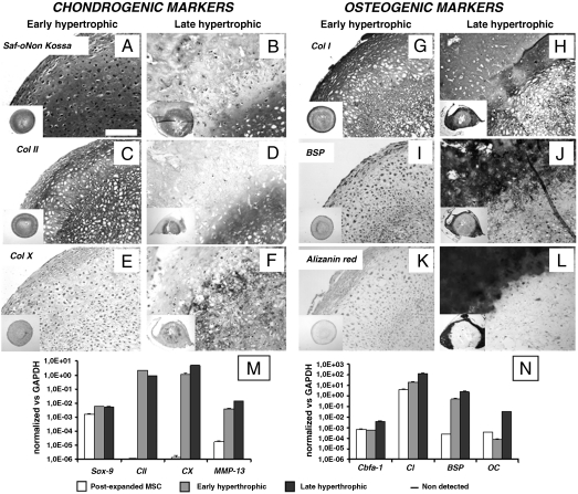
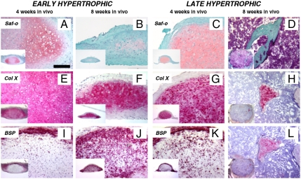
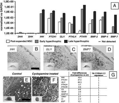
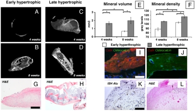
Mesenchymal stem/stromal cells (MSC) are typically used to generate bone tissue by a process resembling intramembranous ossification, i.e., by direct osteoblastic differentiation. However, most bones develop by endochondral ossification, i.e., via remodeling of hypertrophic cartilaginous templates. To date, endochondral bone formation has not been reproduced using human, clinically compliant cell sources. Here, we aimed at engineering tissues from bone marrow-derived, adult human MSC with an intrinsic capacity to undergo endochondral ossification. By analogy to embryonic limb development, we hypothesized that successful execution of the endochondral program depends on the initial formation of hypertrophic cartilaginous templates. Human MSC, subcutaneously implanted into nude mice at various stages of chondrogenic differentiation, formed bone trabeculae only when they had developed in vitro hypertrophic tissue structures. Advanced maturation in vitro resulted in accelerated formation of larger bony tissues. The underlying morphogenetic process was structurally and molecularly similar to the temporal and spatial progression of limb bone development in embryos. In particular, Indian hedgehog signaling was activated at early stages and required for the in vitro formation of hypertrophic cartilage. Subsequent development of a bony collar in vivo was followed by vascularization, osteoclastic resorption of the cartilage template, and appearance of hematopoietic foci. This study reveals the capacity of human MSC to generate bone tissue via an endochondral program and provides a valid model to study mechanisms governing bone development. Most importantly, this process could generate advanced grafts for bone regeneration by invoking a "developmental engineering" paradigm.


Conflict of interest statement

The authors declare no conflict of interest.

Figures

Fig. 1.
Fig. 1.
In vitro maturation of hypertrophic cartilage tissues engineered from human adult MSC. In vitro culture conditions determined the composition and structure of the tissues generated. (A, C, E, and G) Early hypertrophic samples displayed a cartilaginous ECM rich in GAG and Col II with deposition of Col X and Col I in defined regions. (I and K) In the periphery of early hypertrophic samples, low BSP levels were detected, but no calcium was deposited. (B, D, F, H, J, and L) Late hypertrophic samples underwent further maturation in vitro and developed two distinct regions: an inner hypertrophic core (B, D, and F) rich in GAG, Col II, and Col X, and an outer mineralized rim (B, H, J, and L) with a high mineral content, Col I, and BSP. All pictures were taken at the same magnification. (Scale bar: 200 μm.) The insets display low magnification overviews of the entire tissues. (M and N) Quantitative real-time RT-PCR demonstrated an up-regulation of hypertrophic (Col X and MMP-13) and osteogenic (cbfa-1, OC, BSP) markers when comparing late with early hypertrophic tissues. Postexpanded MSC remained in an undifferentiated state but expressed both SOX-9 and Cbfa-1 in combination with high type I collagen and low type II collagen levels.
Fig. 2.
Fig. 2.
Development of the hypertrophic cartilage tissues following in vivo implantation. The differentiation of cartilaginous constructs in vivo progressed according to their stage of in vitro maturation. (A, E, and I) Four weeks after implantation, early hypertrophic samples had differentiated further toward hypertrophy, displaying larger lacunae, Col X accumulation, and initiated BSP deposition in the outer rim. (B, F, and J) Eight weeks after implantation, early hypertrophic samples had differentiated even further. This was evidenced by a decrease in GAG accumulation, while Col X was maintained and BSP had also been deposited within the cartilaginous core. (C, G, and K) After 4 weeks, late hypertrophic specimens had undergone more intense remodeling, such that GAG and Col X levels were reduced, while BSP had already been deposited within the cartilaginous core. (D, H, and L) After 8 weeks, the cartilaginous template was almost completely resorbed: Bone structures substituted the GAG positive areas in the central region, while Col X and BSP positive areas were restricted to scattered islands. All the pictures were taken at the same magnification. (Scale bar: 200 μm.)
Fig. 3.
Fig. 3.
In vivo remodeling and vascularization of late hypertrophic cartilage implants. (A) The observed remodeling resembles the temporal and spatial changes indicative of ongoing endochondral ossification. Hypertrophic chondrocytes located in the bony collar (BC) synthesized MMP13, which is known to prepare the ECM for vascular invasion during endochondral ossification. (B and C) Newly formed vessels, identified by CD31+ endothelial cells, penetrated the outer matrix and reached the inner core in close proximity to the cartilaginous areas undergoing remodeling (arrows). (D and E) The cartilaginous regions were colonized by TRAP-positive cells synthesizing MMP9. (F) These regions were also positive for the DIPEN, which is produced by MMP-mediated cleavage of aggrecan. (Scale bar: 100 μm.)
Fig. 4.
Fig. 4.
Activation of signaling pathways involved in endochondral bone formation in embryos. Signaling pathways typically involved in endochondral ossification were activated in the engineered samples. (A) Real-time RT-PCR analysis indicated that MSC cultured under hypertrophic conditions up-regulated the expression of genes in the IHH signaling pathway (involving IHH, GLI1, and PTCH1), BMPs and parathyroid hormone-related protein signaling (PTHLH, PTHR1). Note that all fold changes in transcript levels are shown in logarithmic scale. (BD) Four weeks after implantation, the expression of representative genes was assessed by ISH (IHH, GLI1, and BMP7). (EG) Functional inhibition of the IHH pathway by cyclopamine treatment significantly reduced the expression of genes involved in IHH signaling (IHH, GLI1, PTCH1), PTH signaling (PTHLH, PTHR1), as well as chondrogenic/hypertrophic genes (Col II, VEGF), and osteogenic genes (Cbfa-1, Col I, BSP). Cyclopamine also blocked the differentiation and maturation of the cartilaginous templates in vitro, as assessed by Safranin-O stain. [Scale bar: 200 μm (BD).] [Scale bar: 400 μm (E and F)].
Fig. 5.
Fig. 5.
Morphometric analysis of the engineered bone tissue. (AD) Three-dimensional μCT reconstructions and (E and F) quantitative histomorphometric data (n = 4) of mineral volume and density indicate higher bone quantity and more advanced maturation of late hypertrophic samples (* indicates significant differences; p < 0.01). (G and H) Trabecular-like structures were found both in the outer bony collar and in the inner core of late, but not early, hypertrophic samples. (Scale bar: 200 μm.) (I and J) Fluorescence characterization for Col X (red) and osteocalcin (green) demonstrated the presence of mature lamellar bone only in late hypertrophic samples. (Scale bar: 50 μm.) (K and L) ISH to detect human Alu repeat sequences and hematoxylin/eosin staining of serial sections indicate that cells derived from the human adult MSC participated in the endochondral ossification process. (Scale bar: 100 μm.)

Similar articles

Cited by

References

    1. Kronenberg HM. Developmental regulation of the growth plate. Nature. 2003;423:332–336. - PubMed
    1. Noonan KJ, Hunziker EB, Nessler J, Buckwalter JA. Changes in cell, matrix compartment, and fibrillar collagen volumes between growth-plate zones. J Orthop Res. 1998;16:500–508. - PubMed
    1. Bianco P, Cancedda FD, Riminucci M, Cancedda R. Bone formation via cartilage models: The “borderline” chondrocyte. Matrix Biol. 1998;17:185–192. - PubMed
    1. Meijer GJ, de Bruijn JD, Koole R, van Blitterswijk CA. Cell-based bone tissue engineering. PLoS Med. 2007;4:e9. doi: 10.1371/journal.pmed.0040009. - DOI - PMC - PubMed
    1. Ashton BA, et al. Formation of bone and cartilage by marrow stromal cells in diffusion chambers in vivo. Clin Orthop Relat Res. 1980;151:294–307. - PubMed

Publication types

LinkOut - more resources