. 2010 Apr 20;107(16):7281-6.
doi: 10.1073/pnas.1002951107. Epub 2010 Apr 6.

Protein structural dynamics in solution unveiled via 100-ps time-resolved x-ray scattering

Affiliations

Protein structural dynamics in solution unveiled via 100-ps time-resolved x-ray scattering

Hyun Sun Cho et al. Proc Natl Acad Sci U S A. .

Abstract

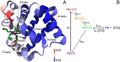
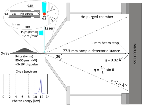
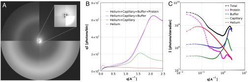
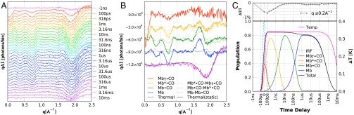
We have developed a time-resolved x-ray scattering diffractometer capable of probing structural dynamics of proteins in solution with 100-ps time resolution. This diffractometer, developed on the ID14B BioCARS (Consortium for Advanced Radiation Sources) beamline at the Advanced Photon Source, records x-ray scattering snapshots over a broad range of q spanning 0.02-2.5 A(-1), thereby providing simultaneous coverage of the small-angle x-ray scattering (SAXS) and wide-angle x-ray scattering (WAXS) regions. To demonstrate its capabilities, we have tracked structural changes in myoglobin as it undergoes a photolysis-induced transition from its carbon monoxy form (MbCO) to its deoxy form (Mb). Though the differences between the MbCO and Mb crystal structures are small (rmsd < 0.2 A), time-resolved x-ray scattering differences recorded over 8 decades of time from 100 ps to 10 ms are rich in structure, illustrating the sensitivity of this technique. A strong, negative-going feature in the SAXS region appears promptly and corresponds to a sudden > 22 A(3) volume expansion of the protein. The ensuing conformational relaxation causes the protein to contract to a volume approximately 2 A(3) larger than MbCO within approximately 10 ns. On the timescale for CO escape from the primary docking site, another change in the SAXS/WAXS fingerprint appears, demonstrating sensitivity to the location of the dissociated CO. Global analysis of the SAXS/WAXS patterns recovered time-independent scattering fingerprints for four intermediate states of Mb. These SAXS/WAXS fingerprints provide stringent constraints for putative models of conformational states and structural transitions between them.


Conflict of interest statement

The authors declare no conflict of interest.

Figures

Fig. 1.
Fig. 1.
(A) Structural differences between MbCO and Mb. The heme, distal His64, and proximal His93 are rendered as licorice for both MbCO (magenta) and Mb (green). The backbone is rendered as ribbon and color coded according to the rmsd between these two structures. Structural alignment and rendering were carried out using the software VMD (33). (B) Five-state model for describing protein and ligand dynamics. Photolysis of MbCO produces Mb•CO, an unrelaxed state with CO harbored in the primary docking site near the binding site. Tertiary structure relaxation is modeled in two steps, and produces Mb•CO with CO still harbored in the primary docking site. From this site, CO can either rebind geminately to recover MbCO, or can escape into the surrounding solvent or migrate into other internal cavities of the protein to produce Mb, which corresponds to deoxy myoglobin. Bimolecular CO binding regenerates the starting state, MbCO, and completes the fully reversible cycle. The color code for this five-state model is maintained throughout this manuscript.
Fig. 2.
Fig. 2.
Sample-detector geometry. The 12 keV x-ray energy, 177.3-mm sample-detector distance, 1-mm beamstop diameter, and ∼165-mm MarCCD detector dimension defines the range of q accessible (∼0.02 to ∼2.5 -1). The x-ray pulse (∼94 ps FWHM; ∼6 μJ; 12 keV) is focused to 80 × 50 μm (FWHM) in the plane of the sample capillary (vertical dimension is smaller). Its asymmetric spectrum is sharply peaked at 12 keV and has a bandwidth of ∼320 eV (2.7% FWHM). The circularly polarized laser pulse (∼35 ps FWHM; 115 μJ; 480 nm) is focused to a 120 × 600 μm (FWHM) elliptical spot on the capillary (red circle) with the long axis aligned along the x-ray beam direction (power density ∼2.1 mJ/mm2). It is set to arrive in coincidence (t = 0) with the x-ray pulse. A 1.5-mm hole was bored into the Mylar film in front of the He-purged chamber (see expanded view).
Fig. 3.
Fig. 3.
(A) Composite 2D image of x-ray scattering from helium, capillary, buffer, and protein solution (quadrants 1–4, respectively). An enlarged view of the SAXS region is shown in the corner of quadrant 1. The protein concentration was ∼50 mg/mL. (B) Angular integration of experimental scattering images in A with the detector pixels binned into annular rings spaced by 0.01 -1. (C) Decomposition of the protein solution scattering pattern into its respective contributions. Note the log–log scale.
Fig. 4.
Fig. 4.
(A) Time-resolved SAXS/WAXS differences. For clarity, the curves are color-coded according to the model in Fig. 1B and offset from one another. (B) Time-independent scattering fingerprints extracted from global analysis of the time-resolved scattering data in A. Scattering differences between each intermediate state and the ground state (MbCO) are plotted as solid lines, whereas differences between each state and the state that precedes it are plotted as dotted lines (three-point smoothing has been applied to the dotted lines). For clarity, the curves are offset vertically from one another. A scaled thermal signal from static measurements (gray) is plotted on top of the thermal signal recovered from global analysis of the time-resolved scattering patterns. (C, Upper) Time-dependence of the integrated SAXS signal. (Lower) Time-dependent population of states in B. The dashed line labeled IRF (cyan) represents the instrument response function (convolution of the laser and x-ray pulses).

References

    1. Schotte F, et al. Watching a protein as it functions with 150-ps time-resolved x-ray crystallography. Science. 2003;300:1944–1947. - PubMed
    1. Schotte F, Soman J, Olson JS, Wulff M, Anfinrud PA. Picosecond time-resolved x-ray crystallography: Probing protein function in real time. J Struct Biol. 2004;147:235–246. - PubMed
    1. Haurowitz F. The Equilibrium between hemoglobin and oxygen. H-S Z Physiol Chem. 1938;254:266–272.
    1. Gibson QH. An apparatus for flash photolysis and its application to the reactions of myoglobin with gases. J Physiol. 1956;134(1):112–122. - PMC - PubMed
    1. Hofrichter J, Sommer JH, Henry ER, Eaton WA. Nanosecond absorption spectroscopy of hemoglobin: Elementary processes in kinetic cooperativity. Proc Natl Acad Sci USA. 1983;80:2235–2239. - PMC - PubMed

Publication types

LinkOut - more resources