. 2010 May 1;70(9):3537-46.
doi: 10.1158/0008-5472.CAN-09-4290. Epub 2010 Apr 20.

Requirement of the NF-kappaB subunit p65/RelA for K-Ras-induced lung tumorigenesis

Affiliations

Requirement of the NF-kappaB subunit p65/RelA for K-Ras-induced lung tumorigenesis

Daniela S Bassères et al. Cancer Res. .

Abstract

K-Ras-induced lung cancer is a very common disease, for which there are currently no effective therapies. Because therapy directly targeting the activity of oncogenic Ras has been unsuccessful, a different approach for novel therapy design is to identify critical Ras downstream oncogenic targets. Given that oncogenic Ras proteins activate the transcription factor NF-kappaB, and the importance of NF-kappaB in oncogenesis, we hypothesized that NF-kappaB would be an important K-Ras target in lung cancer. To address this hypothesis, we generated a NF-kappaB-EGFP reporter mouse model of K-Ras-induced lung cancer and determined that K-Ras activates NF-kappaB in lung tumors in situ. Furthermore, a mouse model was generated where activation of oncogenic K-Ras in lung cells was coupled with inactivation of the NF-kappaB subunit p65/RelA. In this model, deletion of p65/RelA reduces the number of K-Ras-induced lung tumors both in the presence and in the absence of the tumor suppressor p53. Lung tumors with loss of p65/RelA have higher numbers of apoptotic cells, reduced spread, and lower grade. Using lung cell lines expressing oncogenic K-Ras, we show that NF-kappaB is activated in these cells in a K-Ras-dependent manner and that NF-kappaB activation by K-Ras requires inhibitor of kappaB kinase beta (IKKbeta) kinase activity. Taken together, these results show the importance of the NF-kappaB subunit p65/RelA in K-Ras-induced lung transformation and identify IKKbeta as a potential therapeutic target for K-Ras-induced lung cancer.


Figures

Figure 1
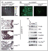
Figure 1. Oncogenic K-Ras activates NF-κB in lung tumors in situ
WT and K-RasG12D/NF-κB mice were analyzed at 19 weeks post-infection. A) In vivo live EGFP fluorescence emission. Color scale indicates the range of fluorescence intensity. B) Laser scanning microscopy of dissected lungs to measure EGFP fluorescence emission. C) Lung tissue sections of WT and K-RasG12D/NF-κB mice were analyzed by GFP immunohistochemistry (positive cells stain in brown). Slides were counterstained with hematoxylin (blue). D) Lung protein lysates from K-RasG12D or K-RasWT mice were submitted to western blotting with the indicated antibodies. Arrows indicate the specific immunodetected bands; additional bands in blots are non-specific.
Figure 2
Figure 2. Loss of p65/RelA reduces formation and spread of K-Ras-induced lung tumors
K-RasG12D/p65WT and K-RasG12D/p65Δ mice were analyzed at 13 weeks post-infection. A) Number of K-Ras-induced neoplastic lesions was determined by counting lesions in Hematoxylin/Eosin stained lung sections as described (see methods). B) PCR to detect cre-mediated recombination of p65/RelA. Bands corresponding to the WT and excised alleles are indicated. C) Immunohistochemistry for p65/RelA (positive cells are brown). Neoplastic lesion and adjacent epithelium are indicated. D) Analysis of lung tumor spread was performed on Hematoxylin/Eosin stained lung sections as described (see methods).
Figure 3
Figure 3. K-RasG12D/p65Δ lung tumors have higher numbers of apoptotic cells
Apoptotic cells were detected by TUNEL staining of lung tissue sections (positive cells are brown).
Figure 4
Figure 4. Loss of p65/RelA reduces formation and grade of K-Ras-induced lung tumors in the absence of p53
K-RasG12D/p53Δ/p65WT and K-RasG12D/p53Δ/p65Δ mice were analyzed at 19 weeks post-infection. A) Number of K-Ras-induced neoplastic lesions was determined by counting lesions in Hematoxylin/Eosin stained lung sections as described (see methods). B) Analysis of lung tumor grade was performed on Hematoxylin/Eosin stained lung sections as described (see methods).
Figure 5
Figure 5. K-Ras-induced NF-κB activation in lung primary cells requires IKKβ activity
A) Nuclear extracts of lung primary SALEB and SAKRAS cells were analyzed by EMSA with a probe containing a canonical NF-κB DNA binding site. Cells were eiter left untreated (UT) or treated for 1h with 20ng/ml TNFα (TNFα), 1uM CmpdA (CmpdA) or 0.02% DMSO (DMSO). B) Expression of NF-κB target genes IL-8, Bcl-XL and MMP-9 was analyzed by real-time quantitative PCR in SALEB and SAKRAS cells treated with 0.02% DMSO (DMSO) or 1uM CmpdA (CmpdA) for 24h. C) Western blotting of total protein lysates from SALEB (SL) or SAKRAS (SK) cells probed with different antibodies as indicated.
Figure 6
Figure 6. K-Ras-induced NF-κB activation in lung cancer cells requires IKKβ expression and activity
A) H358 cells were transiently transfected with siRNA targeted against K-Ras (siK-Ras), IKKβ (siIKKβ), and nontargeting control (siCtrl) and analyzed at 96h post-transfection for NF-κB DNA binding activity by EMSA (left panel) and efficiency of knockdown by Western Blotting (WB, right panel). NF-κB DNA binding complexes as well as antibodies used are indicated. B and C) Dual luciferase reporter assays were performed using an NF-κB-responsive firefly luciferase reporter (3x-κB-Luc, unfilled bars) or an NF-κB-unresponsive reporter (3X-MUTκB-Luc, black-filled bars). For panel B, H358 cells were either transfected with an empty pcDNA3 vector (VC) or with pcDNA3-IKKβ-KM. For panel C, H358 cells were either treated with 0.02% DMSO or treated 1uM CmpdA for 16h. D) DMSO or CmpdA-treated KE67 cells were analyzed by flow cytometry. Forward and side scatter (FS and SS) were used to gate on live cells. The single color histogram represents the number of events recorded in function of the level of GFP fluorescence emission using a logaritmic scale. A threshold for GFP-positive cells was arbitrarily set (linear gate) and the percentage of GFP positive cells is indicated.

Similar articles

Cited by

References

    1. Jemal A, Siegel R, Ward E, Hao Y, Xu J, Thun MJ. Cancer Statistics, 2009. CA Cancer J Clin. 2009;59:225–49. - PubMed
    1. Mills NE, Fishman CL, Rom WN, Dubin N, Jacobson DR. Increased Prevalence of K-ras Oncogene Mutations in Lung Adenocarcinoma. Cancer Res. 1995;55:1444–7. - PubMed
    1. Rodenhuis S, Slebos RJ. Clinical significance of ras oncogene activation in human lung cancer. Cancer Res. 1992;52:2665s–9s. - PubMed
    1. Slebos RJ, Rodenhuis S. The ras gene family in human non-small-cell lung cancer. J Natl Cancer Inst Monogr. 1992:23–9. - PubMed
    1. Graziano SL, Gamble GP, Newman NB, Abbott LZ, Rooney M, Mookherjee S, et al. Prognostic Significance of K-ras Codon 12 Mutations in Patients With Resected Stage I and II Non–Small-Cell Lung Cancer. J Clin Oncol. 1999;17:668. - PubMed

Publication types

MeSH terms