. 2010 May 1;70(9):3483-93.
doi: 10.1158/0008-5472.CAN-09-3107. Epub 2010 Apr 20.

Anti-vascular endothelial growth factor therapies as a novel therapeutic approach to treating neurofibromatosis-related tumors

Affiliations

Anti-vascular endothelial growth factor therapies as a novel therapeutic approach to treating neurofibromatosis-related tumors

Hon Kit Wong et al. Cancer Res. .

Abstract

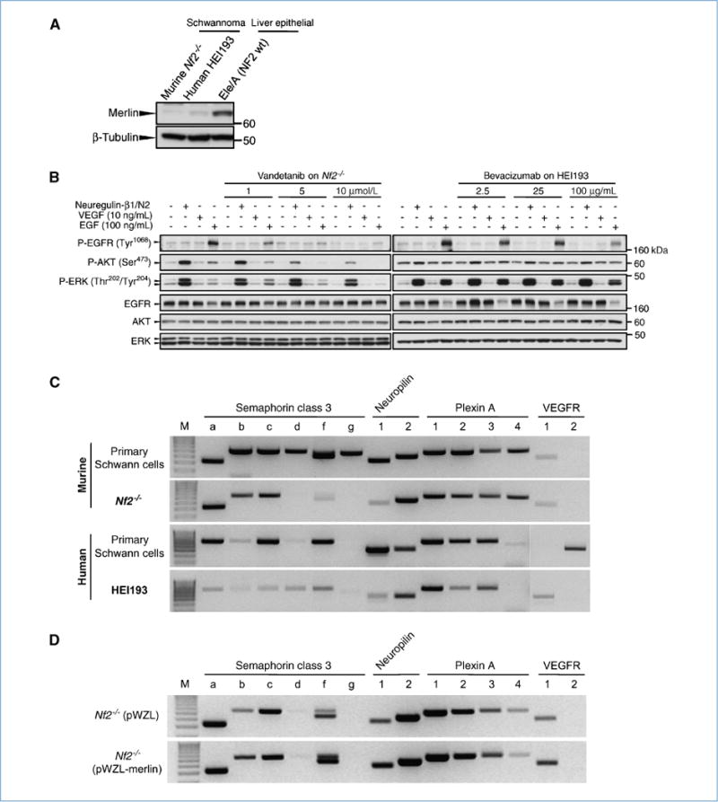
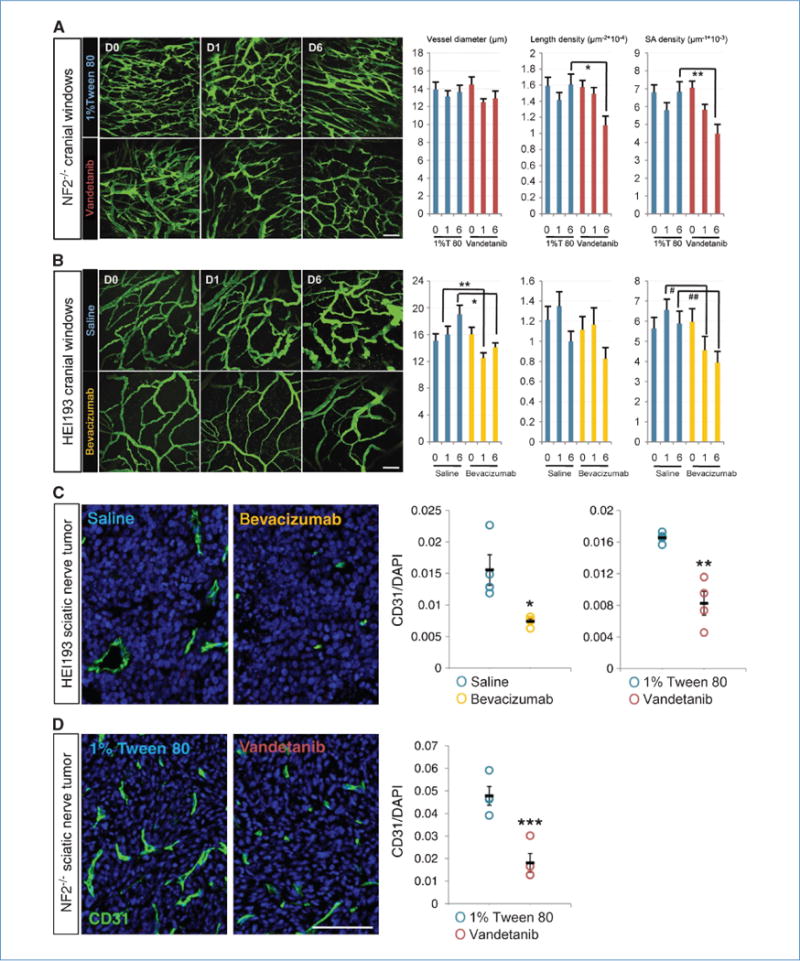
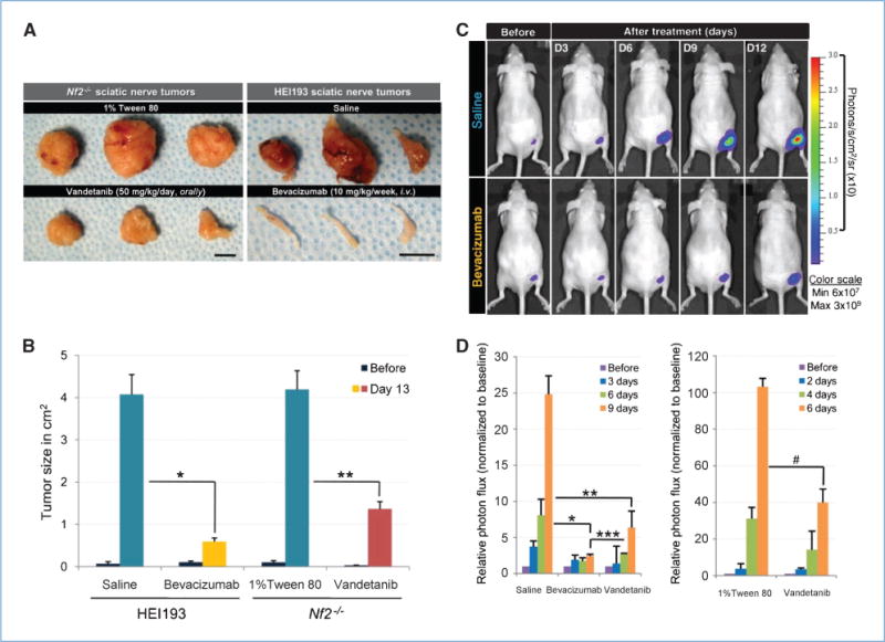
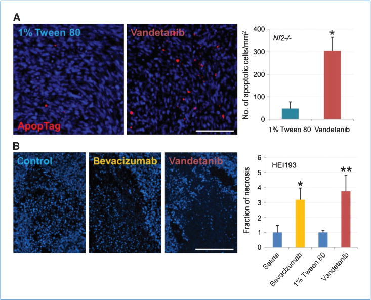
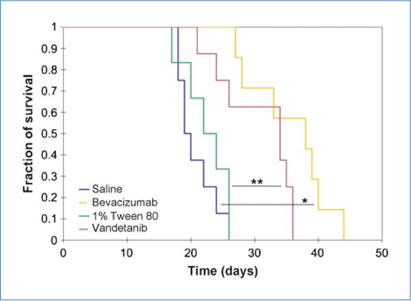
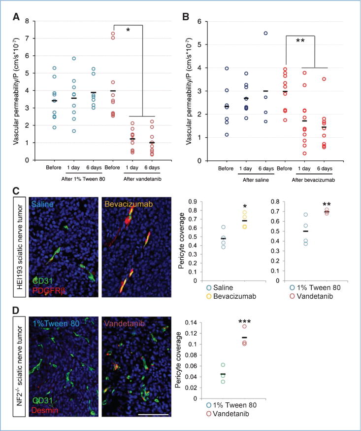
Patients with bilateral vestibular schwannomas associated with neurofibromatosis type 2 (NF2) experience significant morbidity such as complete hearing loss. We have recently shown that treatment with bevacizumab provided tumor stabilization and hearing recovery in a subset of NF2 patients with progressive disease. In the current study, we used two animal models to identify the mechanism of action of anti-vascular endothelial growth factor (VEGF) therapy in schwannomas. The human HEI193 and murine Nf2(-/-) cell lines were implanted between the pia and arachnoid meninges as well as in the sciatic nerve to mimic central and peripheral schwannomas. Mice were treated with bevacizumab (10 mg/kg/wk i.v.) or vandetanib (50 mg/kg/d orally) to block the VEGF pathway. Using intravital and confocal microscopy, together with whole-body imaging, we measured tumor growth delay, survival rate, as well as blood vessel structure and function at regular intervals. In both models, tumor vessel diameter, length/surface area density, and permeability were significantly reduced after treatment. After 2 weeks of treatment, necrosis in HEI193 tumors and apoptosis in Nf2(-/-) tumors were significantly increased, and the tumor growth rate decreased by an average of 50%. The survival of mice bearing intracranial schwannomas was extended by at least 50%. This study shows that anti-VEGF therapy normalizes the vasculature of schwannoma xenografts in nude mice and successfully controls the tumor growth, probably by reestablishing a natural balance between VEGF and semaphorin 3 signaling.


Conflict of interest statement

Disclosure of Potential Conflicts of Interest

S.R. Plotkin: commercial research grant, PTC Therapeutic and Pfizer. R.K. Jain: commercial research grant, AstraZeneca and Dyax; consultant/advisory board, AstraZeneca, Dyax, Enlight, and Millenium; lecture fee from Roche Pharmaceutical. The other authors disclosed no potential conflicts of interest.

Figures

Figure 1
Figure 1
Characterization of a novel schwannoma line and the effect of VEGF inhibitors on these cell lines in vitro. A, wt merlin is below the detection limit both in the newly established murine schwannoma line (Nf2−/−) and in the human HEI193. Liver epithelial cells derived from the Nf2 wt mice served as a merlin-positive control. B, effect of vandetanib and bevacizumab on Nf2−/− and HEI193 signaling. Nf2-deficient or HEI193 schwannoma at density of 2.5 × 10⁶ cells were starved overnight for 12 hours with DMEM and incubated with full medium, recombinant mouse VEGF (10 ng/mL), or human EGF (100 ng/mL) with various concentration of vandetanib or bevacizumab for 15 minutes at 37°C as indicated in the diagram. Vandetanib decreased P-EGFR, P-AKT, and P-ERK in a concentration-dependent manner, whereas bevacizumab did not affect HEI193 signaling unless at very high concentration. C, expression pattern for SEMA3, its receptors, and the VEGF receptors in murine wt Schwann cells and their Nf2−/− counterpart (top) and human Schwann cells compared with HEI193 (bottom). Loss of the Nf2 gene is concurrent with a loss of SEMA3 (b, d, f, and g in murine, all in human), NRP1, and VEGFR1 expression, whereas NRP2 and plexins retain their initial expression. VEGF receptors are not typically expressed by Schwann cells, and oncogenic transformation did not seem to change this feature in vitro. D, reintroduction of the Nf2 gene is accompanied by a reexpression of SEMA3, NRP1, and VEGFR1, supporting the hypothesis of a link between loss of merlin and a change in balance within the angiogenic pathway.
Figure 2
Figure 2
Inhibition of VEGF pathways reduces number and density of schwannoma vessels in brain (A and B) and nerve microenvironment (C and D). Vandetanib reduced vessel length and surface area density of brain Nf2−/− schwannoma without affecting vessel diameter, as shown by two-photon MPLSM in vivo 6 days after treatment. 1% Tween 80, n = 11; vandetanib, n = 12. *, P = 9.11 × 10⁻⁵; **, P = 8 × 10⁻⁴. Bevacizumab, on the other hand, reduced vessel diameter and vessel surface area density of HEI193 as short as 1 day after treatment, maintained up to 6 days without affecting the length density. Saline, n = 7; bevacizumab, n = 8. *, P = 6.6 × 10⁻⁵; **, P = 1.32 × 10⁻⁵; #, P = 9.07 × 10⁻⁵; ##, P = 5 × 10⁻⁴. Scale bars, 100 μm. In sciatic nerve tumor, both vandetanib and bevacizumab reduced CD31-positive fraction of HEI193 schwannoma 6 days after treatment (C), so did vandetanib on Nf2−/− (D). All n = 4. *, P = 0.0428; **, P = 0.0097; ***, P = 0.0023. Scale bar, 100 μm.
Figure 3
Figure 3
Inhibition of VEGF pathways delays schwannoma growth. Both vandetanib and bevacizumab delayed growth of schwannoma in sciatic nerve using either caliper (A and B) or whole-body imaging (C and D) as a tool for size measurement. A, photographs showing images of sciatic nerve tumors 12 days after treatment. Scale bars, 1 cm. B, keeping track of tumor size using caliper. Tumor sizes were measured every 2 days in both controls and the treated, and there was a marked decrease of tumor size 13 days after treatment. Controls, n = 9; vandetanib, n = 8; bevacizumab, n = 3. *, P = 0.0019; **, P = 0.0082. C, representative bioluminescence images showing time-dependent growth of schwannoma in sciatic nerve and growth delay of tumors that have been treated with bevacizumab. D, tumor sizes were expressed as the total number of photon flux. Both vandetanib and bevacizumab significantly reduced HEI193 tumor size, and it seemed that bevacizumab produced a more long-term suppression (left graph). Saline, n = 9; bevacizumab, n = 8; vandetanib, n = 7. *, P = 1.175 × 10⁻⁶; **, P = 5.988 × 10⁻⁵; ***, P = 0.008. Vandetanib had a similar effect on Nf2−/− (right graph). 1% Tween 80, n = 3; vandetanib, n = 4. #, P = 0.0076.
Figure 4
Figure 4
Inhibition of VEGF pathway increased tumor cell death. A, vandetanib increased Nf2−/− cell apoptosis by 6-fold 6 days after treatment, revealed by ApopTag. n = 4 in both control and treated. *, P = 6.946 × 10⁻⁵. B, bevacizumab increased necrotic fraction of tumor instead, there being a 3-fold increase 6 days after treatment. n = 4 in control and the treated. *, P = 0.0119; **, P = 0.0174. Scale bar, 100 μm.
Figure 5
Figure 5
Both vandetanib and bevacizumab markedly extended survival of mice bearing brain schwannoma. Median survival for saline, 19.5 days (n = 8); for bevacizumab, 38 days (n = 7); for 1% Tween 80, 23 days (n = 6); and for vandetanib, 34 days (n = 8). *, P < 0.001; **, P = 0.016, log-rank test.
Figure 6
Figure 6
Inhibition of VEGF pathway improved function and recovered molecular composition of schwannoma vessels. A and B, both vandetanib and bevacizumab reduced vessel leakiness to rho-BSA 24 hours after treatment and the suppression was maintained for up to 6 days of treatment. For 1% Tween 80: before, n = 9; 1 day, n = 9; and 6 days, n = 7; for vandetanib: before, n = 10; 1 day, n = 11; and 6 days, n = 9; for saline: before, n = 8; 1 day, n = 8; and 6 days, n = 4; for bevacizumab: before, n = 9; 1 day, n = 11; and 6 days, n = 9. *, P < 0.001; **, P = 0.01, analyzed by ANOVA Fisher’s F test. C and D, anti-VEGF treatment increased pericyte coverage of schwannoma blood vessels, defined by PDGFRβ in HEI193 (C) and desmin in Nf2−/− (D) at day 6. All n = 4, except G = 3. *, P = 0.0194; **, P = 0.0303; ***, P = 0.0082. Scale bar, 100 μm.

Similar articles

Cited by

References

    1. Giovannini M, Robanus-Maandag E, van der Valk M, et al. Conditional biallelic Nf2 mutation in the mouse promotes manifestations of human neurofibromatosis type 2. Genes Dev. 2000;14:1617–30. - PMC - PubMed
    1. Harner SG, Laws ER., Jr Diagnosis of acoustic neurinoma. Neurosurgery. 1981;9:373–9. - PubMed
    1. Mrugala MM, Batchelor TT, Plotkin SR. Peripheral and cranial nerve sheath tumors. Curr Opin Neurol. 2005;18:604–10. - PubMed
    1. Caye-Thomasen P, Baandrup L, Jacobsen GK, Thomsen J, Stangerup SE. Immunohistochemical demonstration of vascular endothelial growth factor in vestibular schwannomas correlates to tumor growth rate. Laryngoscope. 2003;113:2129–34. - PubMed
    1. Caye-Thomasen P, Werther K, Nalla A, et al. VEGF and VEGF receptor-1 concentration in vestibular schwannoma homogenates correlates to tumor growth rate. Otol Neurotol. 2005;26:98–101. - PubMed

Publication types

MeSH terms