. 2010 May 1;70(9):3667-76.
doi: 10.1158/0008-5472.CAN-09-3647. Epub 2010 Apr 20.

Mechanism of autophagy to apoptosis switch triggered in prostate cancer cells by antitumor cytokine melanoma differentiation-associated gene 7/interleukin-24

Affiliations

Mechanism of autophagy to apoptosis switch triggered in prostate cancer cells by antitumor cytokine melanoma differentiation-associated gene 7/interleukin-24

Sujit K Bhutia et al. Cancer Res. .

Abstract

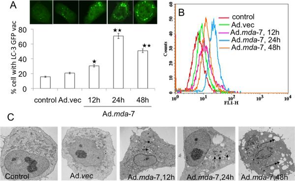
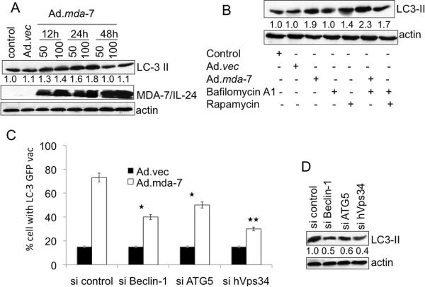
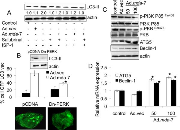
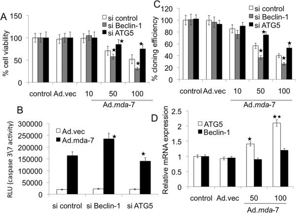
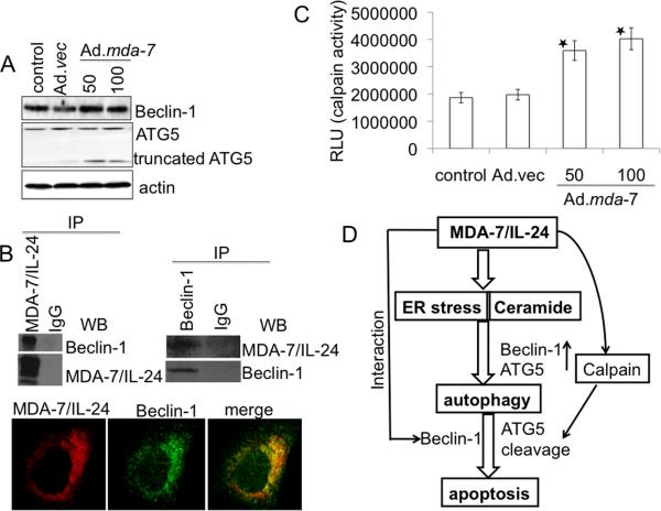
Melanoma differentiation-associated gene 7 (mda-7)/interleukin-24 (IL-24) is a unique member of the IL-10 gene family, which displays a broad range of antitumor properties, including induction of cancer-specific apoptosis. Adenoviral-mediated delivery by Ad.mda-7 invokes an endoplasmic reticulum (ER) stress response that is associated with ceramide production and autophagy in some cancer cells. Here, we report that Ad.mda-7-induced ER stress and ceramide production trigger autophagy in human prostate cancer cells, but not in normal prostate epithelial cells, through a canonical signaling pathway that involves Beclin-1, atg5, and hVps34. Autophagy occurs in cancer cells at early times after Ad.mda-7 infection, but a switch to apoptosis occurs by 48 hours after infection. Inhibiting autophagy with 3-methyladenosine increases Ad.mda-7-induced apoptosis, suggesting that autophagy may be initiated first as a cytoprotective mechanism. Inhibiting apoptosis by overexpression of antiapoptotic proteins Bcl-2 or Bcl-xL increased autophagy after Ad.mda-7 infection. During the apoptotic phase, the MDA-7/IL-24 protein physically interacted with Beclin-1 in a manner that could inhibit Beclin-1 function culminating in apoptosis. Conversely, Ad.mda-7 infection elicited calpain-mediated cleavage of the autophagic protein ATG5 in a manner that could facilitate switch to apoptosis. Our findings reveal novel aspects of the interplay between autophagy and apoptosis in prostate cancer cells that underlie the cytotoxic action of mda-7/IL-24, possibly providing new insights in the development of combinatorial therapies for prostate cancer.


Figures

Fig. 1
Fig. 1. MDA-7/IL-24 induces autophagy in DU-145 cells
A, DU-145 cells were transfected with GFP-LC3 and infected with Ad.mda-7 (100pfu/cell) and after different times localization of LC3 in transfected cells was examined by confocal microscopy (magnification ×100) and autophagosome formation was quantified and data presented as percentage of GFP-LC3–transfected cells with punctate fluorescence to autophagosome formation. A minimum of 100 GFP-LC3–transfected cells were counted. *, P < 0.05; **, P < 0.001, compared with control at the corresponding time. B, The MDC fluorescent intensity of Ad.mda-7-treated DU-145 cells was analyzed by flow cytometry. This result was representative of three different experiments. C, DU-145 cells were infected with 100 pfu/cell of Ad.mda-7 or Ad.vec for different times and cells were fixed and processed for electron microscopy (single arrow, autophagosome; double arrow, empty vacuole; Scale bars: control and others-2μm).
Fig. 2
Fig. 2. MDA-7/IL-24 induces canonical autophagy in DU-145 cells
A, DU-145 cells were infected with Ad.mda-7 or Ad.vec for different times and LC3 expression was analyzed by Western blotting. B, LC3 expression was examined after 24 h treatment with Ad.mda-7 or Ad.vec (100 pfu/cell) in the presence of rapamycin (100 nM), or bafilomycin A1 (100 nM). C, Cells were transfected with the indicated siRNAs and LC3-GFP followed by infection with 100 pfu/cell Ad.mda-7 or Ad.vec and cytoplasmic aggregation of LC3-GFP was determined. A minimum of 100 GFP-LC3–transfected cells were counted. *, P < 0.05; **, P < 0.001, compared with si control. D, LC3-II expression 24 h after administration of the indicated siRNAs and Ad.mda-7 infection (100 pfu/cell) by immunoblot. Densitometry was performed on the original blots, and the ratio of LC3-II/actin in control cells was 1.
Fig. 3
Fig. 3. MDA-7/IL-24-induced autophagy is mediated through ER stress and ceramide production
A, DU-145 cells were infected with 100 pfu/cell of Ad.mda-7 or Ad.vec in the presence of Salubrinal (5 μM) and/or ISP-1 (10 μM) for 24 h followed by analysis of LC3 expression by immunoblotting. Densitometry was performed on the original blots, and the ratio of LC3-II/actin in control cells was 1. B, DU-145 cells were transfected with the indicated plasmids: empty vector control plasmid (pCDNA) or a plasmid expressing dominant negative PERK (Dn-PERK) and GFP-LC3. Cells were analyzed for LC3 aggregation and LC3 expression 24 h after Ad.mda-7 or Ad.vec infection (100 pfu/cell). *, P < 0.05; compared with pCDNA Ad.mda-7 infected cells. C, Evaluation of protein expression: Beclin-1, ATG5, total PKB, p-PKB, total PI3K and p-PI3K using immunoblotting 24 h after Ad.mda-7 or Ad.vec infection (100 pfu/cell). D, DU-145 cells were infected with 100 pfu/cell of Ad.mda-7 or Ad.vec and after 24 h expression of Beclin-1 and ATG5 mRNA was determined using Taqman real-time PCR. Values are the mean ± S.D. of three independent experiments and *, p < 0.05 versus control cells.
Fig. 4
Fig. 4. Relationship between autophagy and apoptosis induced by Ad.mda-7
A, MTT assay after infection with Ad.mda-7 or Ad.vec (different pfu/cell) for 48 h in absence or presence of 3-MA (10 mM for 48 h). B, Caspase-Glo(R) 3/7 assay analyses for caspase-3 expression in DU-145 cells infected with Ad.mda-7 or Ad.vec (100 pfu/cell) for 48 h in absence or presence of 3-MA (10 mM, 48 h). Values are the mean ± S.D. of three independent experiments. *, p < 0.05 versus only Ad.mda-7-treated cells. C, Bcl-2 and Bcl-xL overexpressed DU-145 cells were infected with Ad.mda-7 or Ad.vec (100 pfu/cell) for 24 h and analyzed for LC3 aggregation and LC3 expression by immunoblotting. *, p < 0.05 versus Ad.mda-7-treated DU-Neo cells. Densitometry was performed on the original blots, and the ratio of LC3-II/actin in Ad.mda-7-treated neo cells was 1. D, DU-145 cells were infected with Ad.mda-7 or Ad.vec (100 pfu/cell) for 48 h and stained with MDC and PI followed by flow cytometric analysis. This result was representative of three different experiments.
Fig. 5
Fig. 5. Role of Beclin-1 and ATG5 in Ad.mda-7-induced apoptosis
DU-145 cells were transfected with the indicated siRNAs and cell viability was determined by MTT assay (A), Caspase-Glo(R) 3/7 assay (B) for caspase-3 expression and colony formation assays (C) 48 h after infection with Ad.mda-7 (10, 50 or 100 pfu/cell) or Ad.vec (100 pfu/cell). C, Colony formation assays in monolayer culture. Colonies were fixed, stained and counted (> 50 cells) 2 weeks after plating. D, Expression profile of Beclin-1 and ATG5 at the mRNA level 48 h after Ad.mda-7-infection (50 or 100 pfu/cell) of DU-145 cells (*, P < 0.05; **, P < 0.001, compared with control).
Fig. 6
Fig. 6. Role of Beclin-1 and ATG-5 in Ad.mda-7-induced apoptosis
A, Expression profile of Beclin-1 and ATG5 protein levels 48 h post-infection of DU-145 cells with Ad.mda-7 (50 or 100 pfu/cell) or Ad.vec (100 pfu/cell). B, DU-145 cells were infected with 100 pfu/cell of Ad.vec or Ad.mda-7 for 48 h and immunoprecipitated with anti-MDA-7/IL-24 or anti-Beclin-1 followed by immunoblotting with anti-Beclin-1 or anti-MDA-7/IL-24 antibodies (upper panel). Fluorescent confocal micrographs of DU-145 showing co-immunolocalization of Beclin-1 and MDA-7/IL-24 (lower panel). C, DU-145 cells were infected with Ad.vec (100 pfu/cell) or Ad.mda-7 (50 or 100 pfu/cell) for 48 h followed by calpain assay using calpain-Glo assay. Values reported are mean ± S.D. of three independent experiments. *, p < 0.05 versus control cells. D, Model illustrating the possible molecular mechanism of ER stress- and ceramide-mediated autophagy promoted by MDA-7/IL-24 that switches to apoptosis in prostate cancer cells.

Similar articles

Cited by

References

    1. Jiang H, Lin JJ, Su ZZ, Goldstein NI, Fisher PB. Subtraction hybridization identifies a novel melanoma differentiation associated gene, mda-7, modulated during human melanoma differentiation, growth and progression. Oncogene. 1995;11:2477–86. - PubMed
    1. Sauane M, Gopalkrishnan RV, Sarkar D, et al. MDA-7/IL-24: novel cancer growth suppressing and apoptosis inducing cytokine. Cytokine Growth Factor Rev. 2003;14:35–51. - PubMed
    1. Pestka S, Krause CD, Sarkar D, Walter MR, Shi Y, Fisher PB. Interleukin-10 and related cytokines and receptors. Annu Rev Immunol. 2004;22:929–79. - PubMed
    1. Gupta P, Su ZZ, Lebedeva IV, et al. mda-7/IL-24: Multifunctional cancer-specific apoptosis-inducing cytokine. Pharmacol Ther. 2006;111:596–628. - PMC - PubMed
    1. Fisher PB, Gopalkrishnan RV, Chada S, et al. mda7-7/IL-24, a novel cancer selective apoptosis inducing cytokine gene: from the laboratory into the clinic. Cancer Biol Ther. 2003;2:S23–37. - PubMed

Publication types