. 2010 May 1;70(9):3739-49.
doi: 10.1158/0008-5472.CAN-09-3531. Epub 2010 Apr 20.

Pituitary tumor transforming gene binding factor: a new gene in breast cancer

Affiliations

Pituitary tumor transforming gene binding factor: a new gene in breast cancer

Rachel J Watkins et al. Cancer Res. .

Abstract

Pituitary tumor transforming gene (PTTG) binding factor (PBF; PTTG1IP) is a relatively uncharacterized oncoprotein whose function remains obscure. Because of the presence of putative estrogen response elements (ERE) in its promoter, we assessed PBF regulation by estrogen. PBF mRNA and protein expression were induced by both diethylstilbestrol and 17beta-estradiol in estrogen receptor alpha (ERalpha)-positive MCF-7 cells. Detailed analysis of the PBF promoter showed that the region -399 to -291 relative to the translational start site contains variable repeats of an 18-bp sequence housing a putative ERE half-site (gcccctcGGTCAcgcctc). Sequencing the PBF promoter from 122 normal subjects revealed that subjects may be homozygous or heterozygous for between 1 and 6 repeats of the ERE. Chromatin immunoprecipitation and oligonucleotide pull-down assays revealed ERalpha binding to the PBF promoter. PBF expression was low or absent in normal breast tissue but was highly expressed in breast cancers. Subjects with greater numbers of ERE repeats showed higher PBF mRNA expression, and PBF protein expression positively correlated with ERalpha status. Cell invasion assays revealed that PBF induces invasion through Matrigel, an action that could be abrogated both by siRNA treatment and specific mutation. Furthermore, PBF is a secreted protein, and loss of secretion prevents PBF inducing cell invasion. Given that PBF is a potent transforming gene, we propose that estrogen treatment in postmenopausal women may upregulate PBF expression, leading to PBF secretion and increased cell invasion. Furthermore, the number of ERE half-sites in the PBF promoter may significantly alter the response to estrogen treatment in individual subjects.


Figures

Figure 1
Figure 1
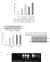
A. PBF mRNA expression was induced maximally at 48 hours by 20 nM diethylstilbestrol (DES) and 17ß-estradiol (EST) in ERα-positive MCF-7 cells compared to vehicle treated cells. ΔCt values from TaqMan RT-PCR with associated standard errors of the mean (SEM) are given below. B. PBF protein expression levels were up-regulated by 20 nM diethylstilbestrol and 17ß-estradiol. Scanning densitometry (N=3 experiments) was used to calculate a ratio of expression compared to β-Actin. Vehicle treatment (V) = 1.0. C. Oestrogen (17ß-estradiol) and anti-oestrogen ICI 182780 treatment of MCF-7 cells at 48 hours. V = vehicle. D. Crosslinking chromatin immunoprecipitation (ChIP) analysis of ERα binding to the human PBF promoter, yielding an expected product of 286 bp. IgG = immunoglobin control; NTC = no template control. *p<0.05; **p<0.01; ***p<0.001.
Figure 2
Figure 2
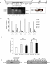
Polymorphism of the PBF promoter. A. A hypervariable region between −399 and −292 upstream of the ATG contains between 1 and 6 repeats of an 18bp motif, which houses a putative consensus ERE half-site (GGTCA). TSS – transcriptional start site. Below - PCR analysis of the PBF promoter, showing negative control (−ve), positive control (+ve; plasmid containing 3 repeats), and 2 individuals with 3 and 5 (3/5) and 3 and 6 (3/6) repeats of the 18 bp motif. L – ladder. An alternative set of primers (Primer Set 2) was used to amplify the PBF promoter region. B. Allele frequencies for 1 to 6 half-site repeats in N = 122 subjects. C. Oestrogen responsiveness of the promoter region −510 to −211 was examined through luciferase assays at 24 and 48 hours post-transfection in MCF-7 cells with 20 nM diethylstilbestrol (DES) and 17ß-estradiol (EST). Data were corrected for Renilla activity and are expressed relative to vehicle treatment. D. Oligonucleotide pull down assay of recombinant human ERα binding to 3 ERE half sites in the human PBF promoter (PBF). NEG = negative control, lacking PBF oligonucleotide. ERE = biotinylated consensus double stranded ERE oligonucleotide (22). PBF Mut = mutated EREs from the human PBF promoter. *p<0.05; **p<0.01.
Figure 3
Figure 3
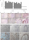
A. Expression of PBF mRNA relative to 18s rRNA (ΔCt values) in 20 TMA samples of breast tumours compared with normal breast. ND – Not detected after 40 cycles of PCR. B. Representative immunohistochemical examination of PBF staining in 1 normal breast sample (US BioMax, Rockville, MD, USA) and 7 tumour samples from TMA sections. Columns 1 to 4 represent the different staining intensities observed, from 0 (absent) to +3 (intense). Values in the bottom right hand corners indicate the percentage of PBF expression observed in the whole section, with original magnifications annotated next to tumour type. C. Representative immunohistochemical examination of PBF staining in 3 normal breast samples (N1-N3; US BioMax, Rockville, MD, USA; 40× original magnification) and 3 tumour samples from TMA sections. T1 - Grade I; T2 – Grade II; T3 – Grade III; all 40× original magnification.
Figure 4
Figure 4
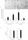
A. PBF over-expression significantly increased the invasiveness of MCF-7 cells after both 24 and 48 hours compared to vector-only (VO). Representative images of invading cells (arrowed) stained with Mayer's Haematoxylin (nuclear, blue) and eosin (cytoplasm, pink) are presented above. **p<0.01. B. MCF-7 cell number following transfection with vector only (VO) or PBF assessed through MTT assays. * p<0.05, N=4.
Figure 5
Figure 5
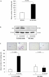
A. 10 nM diethylstilbesterol treatment of 1×10⁵ native MCF-7 cells induced significant invasion through Matrigel at 24 hours. B. siRNA knock-down of PBF. 10 nM DES induced PBF in the presence of a scrambled siRNA control, compared to vehicle treatment. However, 50 nM PBF siRNA yielded 80 to 90 % knock-down of protein, which was not altered by DES treatment. C. Invasion assays carried out in parallel to knock-down experiments. DES = diethylstilbestrol, V = vehicle. Invading cells are arrowed.
Figure 6
Figure 6
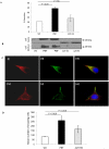
Effect of wild type and mutant PBF overexpression on the secretion and invasiveness of MCF-7 cells. A. The percentage of total PBF secreted into the medium extracted from MCF-7 cells. Mutant Δ29-93 lacks amino acids 29 to 93. B. Western blot of cell medium and whole cell lysate extracted from MCF-7 cells transfected with VO, wild type PBF and the Δ29-93 PBF mutant. PBF constructs were HA-tagged. Whereas WT PBF is detectable in the cell medium, Mutant Δ29-93 is not. C. Immunofluorescent subcellular analysis of MCF-7 cells co-transfected with HA-tagged PBF and chromogranin A-GFP. (i) and (iv) – vesicular PBF-HA expression (red); (ii) and (v) – chromogranin A-GFP expression; (iii) and (vi) – merged image of PBF-HA and chromogranin A-GFP. D. Cell invasion assays. In contrast to WT PBF, Mutant Δ29-93 failed to induce statistically significant cell invasion. Mean values ± SEM are shown. N = 10.

Similar articles

Cited by

References

    1. Allred DC, Medina D. The relevance of mouse models to understanding the development and progression of human breast cancer. J Mammary Gland Biol Neoplasia. 2008;13:279–88. - PubMed
    1. Boelaert K, Tannahill LA, Bulmer JN, Kachilele S, Chan SY, Kim D, et al. A potential role for PTTG/securin in the developing human fetal brain. FASEB J. 2003;17:1631–9. - PubMed
    1. Boelaert K, Smith VE, Stratford AL, Kogai T, Tannahill LA, Watkinson JC, et al. PTTG and PBF repress the human sodium iodide symporter. Oncogene. 2007;26:4344–56. - PubMed
    1. Chien W, Pei L. A novel binding factor facilitates nuclear translocation and transcriptional activation function of the pituitary tumor-transforming gene product. J Biol Chem. 2000;275:19422–7. - PubMed
    1. Mo Z, Zu X, Xie Z, Li W, Ning H, Jiang Y, et al. Antitumor effect of F-PBF(beta-TrCP)-induced targeted PTTG1 degradation in HeLa cells. J Biotechnol. 2009;139:6–11. - PubMed

Publication types

MeSH terms