. 2010 Jul 1;115(26):5418-26.
doi: 10.1182/blood-2009-11-252338. Epub 2010 Apr 20.

Netrin-4 induces lymphangiogenesis in vivo

Affiliations

Netrin-4 induces lymphangiogenesis in vivo

Frederic Larrieu-Lahargue et al. Blood. .

Abstract

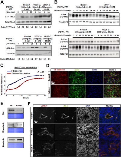
Netrin-4, a laminin-related secreted protein is an axon guidance cue recently shown essential outside of the nervous system, regulating mammary and lung morphogenesis as well as blood vascular development. Here, we show that Netrin-4, at physiologic doses, induces proliferation, migration, adhesion, tube formation and survival of human lymphatic endothelial cells in vitro comparable to well-characterized lymphangiogenic factors fibroblast growth factor-2 (FGF-2), hepatocyte growth factor (HGF), vascular endothelial growth factor-A (VEGF-A), and vascular endothelial growth factor-C (VEGF-C). Netrin-4 stimulates phosphorylation of intracellular signaling components Akt, Erk and S6, and their specific inhibition antagonizes Netrin-4-induced proliferation. Although Netrin receptors Unc5B and neogenin, are expressed by human lymphatic endothelial cells, suppression of either or both does not suppress Netrin-4-promoted in vitro effects. In vivo, Netrin-4 induces growth of lymphatic and blood vessels in the skin of transgenic mice and in breast tumors. Its overexpression in human and mouse mammary carcinoma cancer cells leads to enhanced metastasis. Finally, Netrin-4 stimulates in vitro and in vivo lymphatic permeability by activating small GTPases and Src family kinases/FAK, and down-regulating tight junction proteins. Together, these data provide evidence that Netrin-4 is a lymphangiogenic factor contributing to tumor dissemination and represents a potential target to inhibit metastasis formation.


Figures

Figure 1
Figure 1
Netrin-4 induces HMVEC-dLy proliferation, migration, tube formation and survival. (A) Mitogenic potential of different doses of Netrin-4 on lymphatic dermal human microvascular endothelial cells (HMVEC-dLys) compared with several known lymphangiogenic growth factors (FGF-2 or bFGF, HGF, VEGF-A (VA), VEGF-C (VC), and complete (CM) or basal cell (BM) culture media. Cell number was assessed using dojindo reagent 72 hours after treatment and expressed as fold increase versus control. (B) HMVEC-dLy proliferation under a single dose of Netrin-4 (500 ng/mL), FGF-2 (40 ng/mL), VEGF-A (50 ng/mL), or VEGF-C (300 ng/mL) assessed every 24 hours for 3 days. (C) Chemotactic effects of different doses of Netrin-4 and VEGF-C on HMVEC-dLys in a Boyden chamber assay. (D) HMVEC-dLy adhesion on various matrixes: Fibronectin (FN), Netrin-4, Collagen I (Col. I), and Poly-L-Lysin (PLL) at 10 μg/mL. (E) In vitro tube formation by HMVEC-dLys under different doses of Netrin-4, FGF-2 (bF), HGF, VEGF-A (VA), VEGF-C (VC), or complete media (CM). (F) Inhibition of serum deprivation–induced HMVEC-dLys apoptosis under different doses of Netrin-4, FGF-2, VEGF-A (VA), VEGF-C (VC), and complete media (CM). *P < .05.
Figure 2
Figure 2
Netrin-4 activates intracellular signaling pathways of HMVEC-dLys. Determination of the phosphorylation of Akt (Ser 473; Serine 473, panels A and D, respectively), p42/p44 (Thr 202/Tyr 204; Threonin 202/Tyrosine 204, panels B and E, respectively), and Ribosomal protein S6 (Ser 235/236; Serine 235/236, panels C and F) by Western blotting after Netrin-4 treatment of HMVEC-dLy. Experiments were performed in triplicate.
Figure 3
Figure 3
Netrin-4 overexpression in mouse skin results in an increased lymphatic density. (A) Schematic of the Netrin-4 overexpression system and genotyping of the various mice. Netrin-4 cDNA was inserted into the constitutively active Rosa26 locus downstream of a LoxP-flanked transcriptional stop signal. In presence of a cre recombinase driven by the keratin14 promoter (indicated as Prom.), the stop signal is excised allowing expression of Netrin-4. (B) Expression of the LacZ gene reporter (Rosa26 LacZ/+) in mouse skin keratinocytes in presence of the Keratin14 cre recombinase (K14 cre/+). Scale bars: 200 and 50 μm. (C) Quantification of Netrin-4 mRNA by qRT-PCR in the skin of Netrin-4–overexpressing mice versus littermate controls. (D) Three-week-old skin-overexpressing Netrin-4 mice are viable but hairless, smaller, and redder than controls (scale bar: 1 cm). Pictures were taken on an Olympus IX71 microscope, at 100× and 400× magnification using a DP30BW Olympus camera and the MicroSuite Basic Edition Olympus software. (E) Skin sections of 3-week-old Netrin-4–overexpressing and littermates control mice costained with an anti–LYVE-1 and an antipodoplanin (indicated as podo.) antibody (dotted box represents the enlarged area shown; scale bars: 200 and 20 μm). (F) Quantification of the LYVE1+/podoplanin+ staining density using ImageJ and normalized to the control group. Pictures in panel E are representative of the quantification reported in panel F.
Figure 4
Figure 4
Netrin-4–overexpressing MCF7 tumors have more lymphatic vessels and are more metastatic. (A) Human breast cancer MCF7 cells (WT) stably transduced with a Netrin-4 (Net4), VEGF-C or empty vector (control). Protein expression was determined in the cell supernatant (SUP), the fraction secreted and bound to the cytoplasmic membranes (Mb) or in the total cell lysate (CL) by Western blot using an anti–Netrin-4 or an anti–VEGF-C antibody. (rNet4; Recombinant Netrin-4). All control, Netrin-4, and VEGF-C MCF7 tumor cells also express at similar levels both GFP and luciferase (B) and proliferate in vitro at an identical rate (C; Ctrl; control empty vector, Net4; Netrin-4; scale bars in panel B: 100 μm and 1 cm). (D) Control, Netrin-4, and VEGF-C–overexpressing MCF7 cells injected subcutaneously into the midline of the back of NOS/SCID mice. Tumors were removed after 12 weeks, sectioned and costained with an anti–VEGFR-3/Flt-4 and anti–LYVE-1 antibody (scale bar: 100 μm). (E) VEGFR-3+/LYVE-1+ costaining density was quantified using ImageJ and represented as fold increase over the control-empty vector tumor condition. (F) GFP-positive tumor cells in the lumen of tumor lymphatic vessels stained with LYVE-1 antibody in the Netrin-4–overexpressing tumors (scale bars: 100 and 20 μm). (G) Luciferase-positive tumor cells metastasized into the auxillary lymph nodes of the Netrin-4 and VEGF-C tumor-bearing mice. (H) luciferase-positive lymph nodes contain GFP-positive tumor cells and are surrounded by enlarged LYVE-1 stained vessels in mice bearing Netrin-4 and VEGF-C–overexpressing tumors, but not in control condition (scale bar: 200 μm). 66C14 murine mammary carcinoma line metastasizing via the lymphatic system to lungs were stably transfected with a Netrin-4 encoding or empty-vector and injected into the exposed inguinal right mammary fat pad of Balb/C mice. Netrin-4–overexpressing 66C14 tumor bearing mice die faster than controls (I) and present more metastatic nodules per lung (J; scale bar: 5 mm). Pictures were taken on an Olympus IX71 microscope, at 100×, 200×, and 400× magnification using a DP30BW Olympus camera and the MicroSuite Basic Edition Olympus software. The number of metastatic nodules per lung was quantified by visual inspection (K). *P < .05.
Figure 5
Figure 5
Netrin-4 induces in vitro lymphatic permeability. (A) Induction of GTP-RhoA and Rac1 by Netrin-4, VEGF-A, and VEGF-C treatment of HMVEC-dLys. (B) Stimulation of the phosphorylation of Tyrosine 416 of Src kinase family (SFK, Tyr416) and the Tyrosin 861 but not the Tyrosine 391 of focal adhesion kinase (FAK; Tyr861 and Tyr391) by Netrin-4 (500 ng/mL) and VEGF-C (500 ng/mL). (C) Measurement of the electrical resistance of the cell monolayer over 24 hours using the ECIS system or (D) immunostained using an anti–VE-cadherin antibody to visualize cell junctions (scale bar: 50 μm) of HMVEC-dLys seeded either on Fibronectin or Fibronectin plus Netrin-4. (E) Membrane fraction proteins prepared from HMVEC-dLys seeded as previously mentioned in panels C and D analyzed for ZO-1, VE-cadherin, and beta-catenin expression (equivalent loading assessed by coomassie blue staining). (F) Control, Netrin-4, VEGF-C overexpressing MCF7 tumor sections stained for the cell junction protein ZO-1 or the lymphatic marker LYVE1 (scale bar: 20 μm). Data presented in panels A through E are from 1 experiment and representative of 2 independent experiments. Pictures were taken on an Olympus IX71 microscope, at 400× magnification using a DP30BW Olympus camera and the MicroSuite Basic Edition Olympus software.

Similar articles

Cited by

References

    1. Sleeman JP, Thiele W. Tumor metastasis and the lymphatic vasculature. Int J Cancer. 2009 Jun 30; - PubMed
    1. Conway EM, Collen D, Carmeliet P. Molecular mechanisms of blood vessel growth. Cardiovasc Res. 2001;49(3):507–521. - PubMed
    1. Cirulli V, Yebra M. Netrins: beyond the brain. Nat Rev Mol Cell Biol. 2007;8(4):296–306. - PubMed
    1. Koch M, Murrell JR, Hunter DD, et al. A novel member of the netrin family, beta-netrin, shares homology with the beta chain of laminin: identification, expression, and functional characterization. J Cell Biol. 2000;151(2):221–234. - PMC - PubMed
    1. Lejmi E, Leconte L, Pedron-Mazoyer S, et al. Netrin-4 inhibits angiogenesis via binding to neogenin and recruitment of Unc5B. Proc Natl Acad Sci U S A. 2008;105(34):12491–12496. - PMC - PubMed

Publication types

MeSH terms