. 2010 Jun 24;115(25):5214-21.
doi: 10.1182/blood-2009-12-258822. Epub 2010 Apr 20.

Pegylated arginase I: a potential therapeutic approach in T-ALL

Affiliations

Pegylated arginase I: a potential therapeutic approach in T-ALL

Claudia P Hernandez et al. Blood. .

Abstract

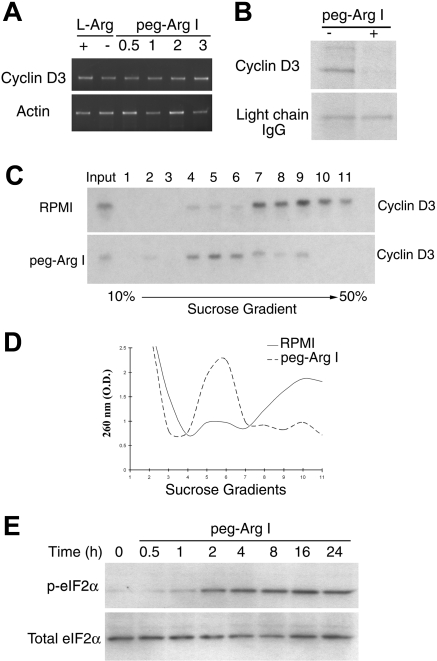
Adult patients with acute lymphoblastic T cell leukemia (T-ALL) have a very poor prognosis and few effective therapeutic options. Therefore, novel therapies that increase the efficacy of the treatments and that prolong T-ALL patient survival are needed. Malignant T cells require high concentrations of nutrients to sustain their increased rate of proliferation. In this study, we determined whether L-Arginine depletion by the pegylated form of the L-Arginine-metabolizing enzyme arginase I (peg-Arg I) impairs the proliferation of malignant T cells. Our results show that peg-Arg I depleted L-Arginine levels in vitro and in vivo. In addition, treatment of malignant T-cell lines with peg-Arg I significantly impaired their proliferation, which correlated with a decreased progression into the cell cycle, followed by the induction of apoptosis. Furthermore, peg-Arg I impaired the expression of cyclin D3, a fundamental protein in T-ALL proliferation, through a global arrest in protein synthesis. Injection of peg-Arg I plus chemotherapy agent Cytarabine prolonged survival in mice bearing T-ALL tumors. This antitumoral effect correlated with an inhibition of T-ALL proliferation in vivo, a decreased expression of cyclin D3, and T-ALL apoptosis. The results suggest the potential benefit of L-Arginine depletion by peg-Arg I in the treatment of T-cell malignancies.


Figures

Figure 1
Figure 1
Peg-Arg I depletes L-Arginine in vitro and in vivo and impairs malignant T-cell proliferation. (A) RPMI medium was treated with peg-Arg I (0.5-4 IU/mL) or PBS and the levels of L-Arginine measured by HPLC after 15 and 30 minutes. (B) Mice were injected with PBS (Control) or peg-Arg I (0.1-1 mg/mouse) and the levels of L-Arginine in the serum were measured every day by HPLC. Five mice were used for each condition. (C) Malignant T-cell lines (1 × 10⁵) were cultured for 72 hours in the presence or the absence of peg-Arg I (2 IU/mL), or the absence of L-Arginine (No-L-Arg), and proliferation determined by MTT. (D) Malignant T-cell lines (1 × 10⁶) were cultured for 24 hours in medium with or without peg-Arg I, after which the cell-cycle progression was determined by propidium iodide. Results are expressed as the percentage of cells in the G0-G1 phase of the cell cycle. Experiments were repeated at least 3 times obtaining similar results.
Figure 2
Figure 2
Peg-Arg I impairs cyclin D3 expression in T-ALL cells. (A) Cyclin D3 expression was determined by Western blot in CCRF-CEM cells cultured with or without peg-Arg I. (B) CCRF-CEM cells (1 × 10⁵) previously transfected with shRNA-coding plasmids against nonrelevant sequences (Control) or cyclin D3 (Cyc D3-shRNA), or transfected with empty plasmids (Empty) were cultured for 72 hours and proliferation tested by MTT. (C) CCRF-CEM cells over-expressing cyclin D3 (Cyclin D3-eGFP), expressing eGFP, or wild-type cells were cultured for 72 hours in RPMI medium (Control) or medium containing peg-Arg I (2 IU/mL), after which proliferation was tested by MTT. Experiments were repeated at least 3 times obtaining similar results.
Figure 3
Figure 3
Peg-Arg I inhibits the expression of cyclin D3 in T-ALL cells through a global arrest in protein synthesis. (A) Cyclin D3 mRNA levels were tested by RT-PCR in CCRF-CEM cells cultured for 24 hours in medium with or without L-Arginine (L-Arg) or in medium containing increasing concentrations of peg-Arg I (0.5-3 IU/mL). (B) Pulse-chase analysis for cyclin D3 in CCRF-CEM cells cultured in the presence or the absence of peg-Arg I (2 IU/mL) for 24 hours. (C) Cyclin D3 mRNA levels were tested in polysomes isolated using sucrose gradients from CCRF-CEM cells cultured in RPMI with or without peg-Arg I for 24 hours. (D) A representative experiment from panel C, in which fractions were tested for absorbance (260 nm) to determine the general distribution of the polysomes. (E) CCRF-CEM cells cultured in medium with peg-Arg I (2 IU/mL) were tested for the expression of phospho-eIF2α by immunoblot. The experiments were repeated a minimum of 3 times obtaining similar results.
Figure 4
Figure 4
Peg-Arg I induces T-ALL apoptosis. (A) CCRF-CEM cells were cultured in medium with or without peg-Arg I and the expression of annexin V was tested after 24, 48, 72, and 96 hours by flow cytometry. (B) Malignant T-cell lines including CCRF-CEM, Molt-3, Molt-4, and Jurkat were cultured as in panel A and tested for the expression of annexin V after 72 hours. Peg-Arg I impaired the mitochondrial membrane potential (C), and induced active caspase-3 expression (D) in CCRF-CEM cells. Values are from 3 similar experiments.
Figure 5
Figure 5
NOD-Scid injected with CCRF-CEM cells develop T-ALL. (A) A representative experiment showing that CCRF-CEM–bearing NOD-Scid mice have an increased accumulation of human CD5+ cells in the spleens after 30 days of tumor injection. (B-C) Spleens from panel A were tested for the expression cyclin D3 and GAPDH by Western blot and for the cell-cycle progression using propidium iodide. The experiments were repeated a minimum of 3 times obtaining similar results.
Figure 6
Figure 6
Peg-Arg I plus Ara-C inhibit the accumulation of T-ALL cell in vivo. (A) Peg-Arg I/Ara-C homogeneously prevents the spleen growth induced by CCRF-CEM cells. Tumor size was tested after 30 days of tumor injection. (B) Survival of CCRF-CEM-bearing mice was established after therapies with PBS (Control; n = 11), peg-Arg I (n = 12), Ara-C (n = 13), or peg-Arg I/Ara-C (n = 13). (C-D) Percentage of human CD5+ cells was established in spleens and peripheral blood after 30 days of tumor injection in mice treated with peg-Arg I/Ara-C (n = 10) or PBS (n = 10).
Figure 7
Figure 7
Peg-Arg I plus Ara-C impairs malignant T-cell proliferation and induces tumor cell apoptosis in vivo. (A-B) Proliferation of T-ALL cells was tested in vivo in CCRF-CEM-bearing mice treated with peg-Arg I/Ara-C (n = 10) or PBS (n = 10) by measuring the uptake of BrdU, as described in “Cell-cycle progression and proliferation.” (C) A representative experiment showing the expression of cyclin D3 and GAPDH in CD5+ cells sorted from individual tumor-bearing mice treated with PBS (n = 3) or peg-Arg I plus Ara-C (n = 5). The experiment was repeated a minimum of 3 times obtaining similar results. (D) Annexin V expression was established in gated CD5+ cells in the spleens from tumor-bearing mice treated with peg-Arg I/Ara-C (n = 10) or PBS (n = 10) after 30 days of tumor injection.

Similar articles

Cited by

References

    1. Pui CH, Evans WE. Acute lymphoblastic leukemia. N Engl J Med. 1998;339(9):605–615. - PubMed
    1. Thomas X, Boiron JM, Huguet F, et al. Outcome of treatment in adults with acute lymphoblastic leukemia: analysis of the LALA-94 trial. J Clin Oncol. 2004;22(20):4075–4086. - PubMed
    1. Laport GF, Larson RA. Treatment of adult acute lymphoblastic leukemia. Semin Oncol. 1997;24(1):70–82. - PubMed
    1. Bronte V, Serafini P, Mazzoni A, Segal DM, Zanovello P. L-arginine metabolism in myeloid cells controls T-lymphocyte functions. Trends Immunol. 2003;24(6):302–306. - PubMed
    1. Morris SM., Jr Recent advances in arginine metabolism. Curr Opin Clin Nutr Metab Care. 2004;7(1):45–51. - PubMed

Publication types

MeSH terms