. 2010 May;120(5):1429-40.
doi: 10.1172/JCI40659. Epub 2010 Apr 19.

Sphingosine kinase 1 and sphingosine-1-phosphate receptor 2 are vital to recovery from anaphylactic shock in mice

Affiliations

Sphingosine kinase 1 and sphingosine-1-phosphate receptor 2 are vital to recovery from anaphylactic shock in mice

Ana Olivera et al. J Clin Invest. 2010 May.

Abstract

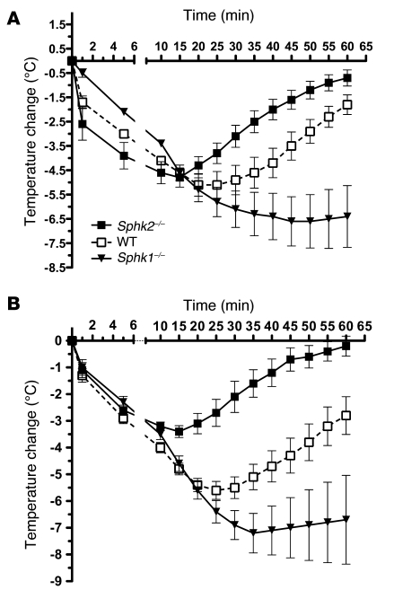
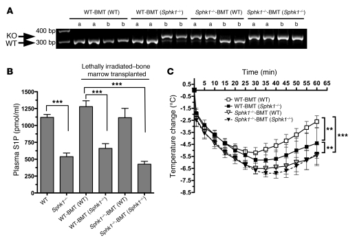
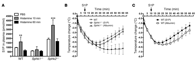
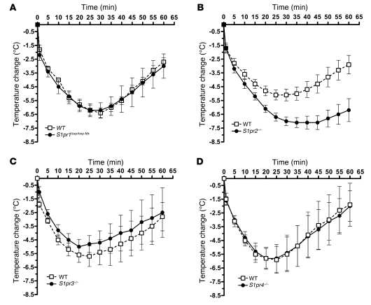
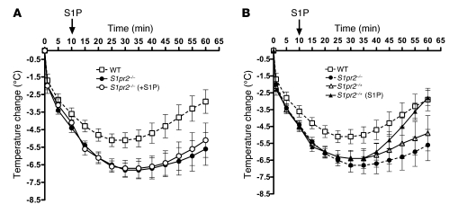
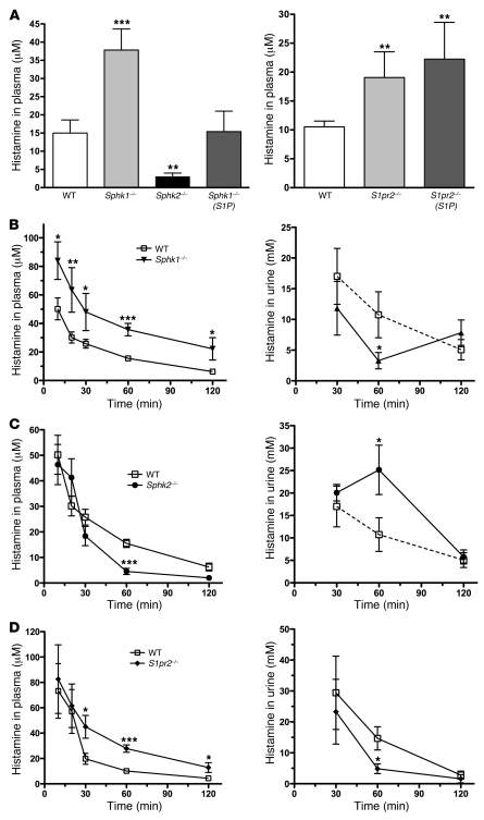
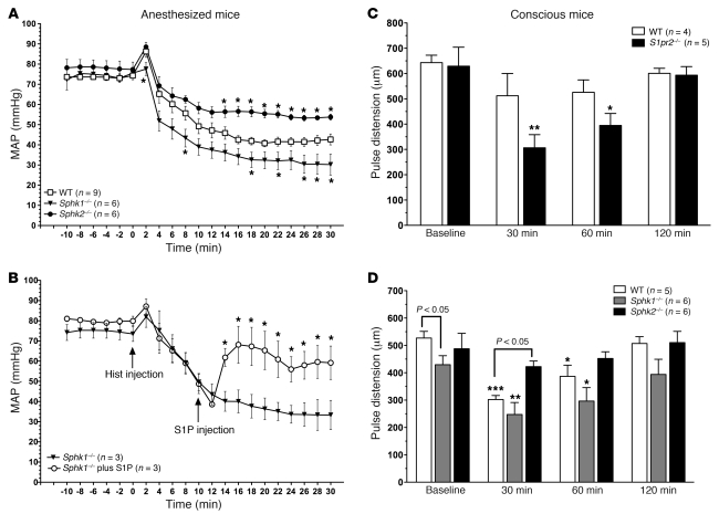
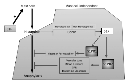
Sphingosine kinase 1 (SphK1) and SphK2 are ubiquitous enzymes that generate sphingosine-1-phosphate (S1P), a ligand for a family of G protein-coupled receptors (S1PR1-S1PR5) with important functions in the vascular and immune systems. Here we explore the role of these kinases and receptors in recovery from anaphylaxis in mice. We found that Sphk2-/- mice had a rapid recovery from anaphylaxis. In contrast, Sphk1-/- mice showed poor recovery from anaphylaxis and delayed histamine clearance. Injection of S1P into Sphk1-/- mice increased histamine clearance and promoted recovery from anaphylaxis. Adoptive cell transfer experiments demonstrated that SphK1 activity was required in both the hematopoietic and nonhematopoietic compartments for recovery from anaphylaxis. Mice lacking the S1P receptor S1PR2 also showed a delay in plasma histamine clearance and a poor recovery from anaphylaxis. However, S1P did not promote the recovery of S1pr2-/- mice from anaphylaxis, whereas S1pr2+/- mice showed partial recovery. Unlike Sphk2-/- mice, Sphk1-/- and S1pr2-/- mice had severe hypotension during anaphylaxis. Thus, SphK1-produced S1P regulates blood pressure, histamine clearance, and recovery from anaphylaxis in a manner that involves S1PR2. This suggests that specific S1PR2 agonists may serve to counteract the vasodilation associated with anaphylactic shock.


Figures

Figure 1
Figure 1. Differences in the recovery from anaphylaxis between Sphk1–/– and Sphk2–/– mice are mast cell–independent.
(A) WT, Sphk1–/–, and Sphk2–/– mice were injected (i.v.) with DNP-specific IgE (3 μg). After 24 hours, mice were challenged with Ag (250 μg DNP36-HSA) to induce systemic anaphylaxis. Body temperature changes during the anaphylactic shock induced by Ag were measured as described in Methods. Mice (n = 12/group) were euthanized after 60 minutes. The gap in the x axis at 6 minutes denotes a change in scale. (B) Anaphylaxis was induced by injecting histamine (5 μmol) into WT, Sphk1–/–, and Sphk2–/– mice (n = 7–10/group). This concentration of histamine showed a similar anaphylactic response as that induced by IgE/Ag in WT mice. The delayed and rapid recoveries of Sphk1–/– and Sphk2–/– mice, respectively, were maintained. Body temperature was monitored as described in Methods.
Figure 2
Figure 2. Involvement of SphK1, in the hematopoietic and nonhematopoietic compartment, in the intensity and recovery from anaphylaxis.
Bone marrow cells from WT or Sphk1–/– mice were adoptively transferred into lethally irradiated WT or Sphk1–/– recipient mice. After engraftment of donor bone marrow cells, mice were subjected to histamine-induced anaphylaxis. (A) A representative figure of PCR-based genotyping results from DNA extracted from tail samples (a) or whole blood (b) of recipient mice showing appropriate engraftment. WT mice showed a 300-bp fragment, whereas Sphk1–/– mice showed a 350-bp fragment. (B) Plasma S1P levels of mice in A were measured as previously described (15). ***P < 0.001, determined by Student’s t test. (C) Body temperature measurements of the lethally irradiated, bone marrow–transplanted mice after a histamine bolus. WT-BMT(WT) mice are lethally irradiated WT recipients reconstituted with donor WT bone marrow cells (n = 13); WT-BMT(Sphk1–/–) mice are WT recipients reconstituted with donor Sphk1–/– bone marrow cells (n = 8); Sphk1–/–-BMT(WT) mice are Sphk1–/– recipients reconstituted with donor WT bone marrow cells (n = 12); and Sphk1–/–-BMT(Sphk1–/–) mice are Sphk1–/– recipients reconstituted with donor Sphk1–/– bone marrow cells (n = 6). **P < 0.01, ***P < 0.001, determined using 2-way ANOVA. Blood analysis in all the lethally-irradiated, bone marrow–transplanted mice showed cell counts similar to those of WT mice, indicating successful engrafting. The blood cell counts present in all chimeras were as follows: lymphocytes, ≥4 × 10⁶/ml; rbc, ~10–11 × 106/μl, platelets, ~2.4–3.8 × 10⁷/ml; monocytes, ~0.5–1.3 × 10⁶/ml; and polymorphonucleated cells, ~1.5–2.6 × 10⁶/ml.
Figure 3
Figure 3. Histamine induces SphK1-dependent increases in plasma S1P levels, and an acute administration of S1P increases the rate of recovery from anaphylactic shock.
(A) WT, Sphk1–/–, and Sphk2–/– mice were injected (i.v.) with histamine (5 μmol) and euthanized after 10 or 60 minutes. Blood was extracted by cardiac puncture and S1P levels were determined. **P < 0.01, ***P < 0.001, determined by Student’s t test. (B and C) Anaphylaxis was induced in Sphk1–/– mice or WT mice by histamine (5 μmol), and after 10 minutes, 500 μM S1P or vehicle (mouse albumin in saline) was administered (i.v.). Body temperature measurements were as described in Methods. (Sphk1–/– [albumin], n = 8; Sphk1–/– [S1P], n = 7; WT [albumin], n = 5; WT [S1P], n = 7).
Figure 4
Figure 4. The S1PR2 receptor controls the duration of anaphylaxis.
(A) S1pr1loxp/loxp-Mx (n = 4), (B) S1pr2–/– (n = 8), (C) S1pr3–/– (n = 4), and (D) S1pr4–/– (n = 4) mice and their respective WT counterparts were subjected to anaphylaxis induced by histamine (5 μmol). The WT counterpart for S1pr1loxp/loxp-Mx mice are S1pr1loxp/loxp mice, as described in Methods. Body temperature measurements were performed as described in Methods.
Figure 5
Figure 5. Administration of S1P does not increase the rate of recovery from anaphylaxis for S1pr2–/– mice.
(A) S1pr2–/– and (B) S1pr2+/– mice were subjected to anaphylaxis by histamine injection (5 μmol). Ten minutes after the histamine bolus, 500 μM S1P or vehicle were administered, and body temperature was monitored as described in Methods. n ≥ 8 in all groups.
Figure 6
Figure 6. Clearance of plasma histamine and excretion are impaired in Sphk1–/– and S1pr2–/– mice but enhanced in Sphk2–/– mice.
(A) Histamine (5 μmol) was injected into the indicated mice, and after 60 minutes, mice were euthanized and blood was drawn. As indicated, 500 μM S1P was administered to some mice 10 minutes after induction of anaphylaxis. Plasma histamine levels were determined for all genotypes and treatments as indicated. (BD) Kinetics of plasma histamine clearance (left panels) and its appearance in urine (right panels) for (B) Sphk1–/–, (C) Sphk2–/–, and (D) S1pr2–/– mice and their corresponding WT counterparts (n = 4–7). Blood was drawn (~2 μl) from each mouse from the tail vein at the indicated times. Urine was collected (when possible) at the indicated times. Histamine was measured by competitive ELISA. *P < 0.05, **P < 0.01, ***P < 0.001, determined by Student’s t test.
Figure 7
Figure 7. Loss of blood pressure is more pronounced in Sphk1–/– and S1pr2–/– mice when compared with WT mice but is less severe in Sphk2–/– mice.
(A) MAP before (10 minutes) and after (30 minutes) histamine injection (5 μmol; at time 0) in isoflurane-anesthetized WT (n = 9), Sphk1–/– (n = 6), and Sphk2–/– mice (n = 6). Graph lines connect mean values of MAP at 2-minute intervals. *P < 0.05, between WT and Sphk–/– mice, determined by Student’s t test. (B) MAP of anesthetized Sphk1–/– mice after histamine injection alone or in combination with S1P treatment (10 minutes after histamine bolus as indicated). The effect of S1P on MAP in the WT mice under anaphylaxis was about half of that seen in the Sphk1–/– mice (data not shown). (C and D) Blood flow in conscious mice determined by pulse distension measurements in (D) WT (n = 6), Sphk1–/– (n = 6), and Sphk2–/– (n = 6) mice or (C) S1pr2–/– (n = 6) and their WT (S1pr2+/+) counterparts (n = 4) at baseline and at 30, 60, and 120 minutes after histamine bolus. *P < 0.05, **P < 0.01, ***P < 0.001, compared with baseline, determined by a Student’s t test.
Figure 8
Figure 8. Proposed model for the dual actions of S1P in the onset of and recovery from anaphylaxis.
While S1P appears to enhance signaling and function of mast cells, which are initiators of the allergic response, it alleviates the symptoms of the anaphylactic shock, acting in a mast cell–independent manner. Histamine released from mast cells activates SphK1 to produce S1P. Although SphK1 in the hematopoietic compartment contributes to the recovery from anaphylaxis, SphK1 in a nonhematopoietic compartment is essential for this function. S1P produced by SphK1 activates S1PR2 receptors, which aid in the clearance of histamine to terminate anaphylaxis. The increased clearance of histamine mediated by S1PR2 appears to be hemodynamic in nature, i.e., a consequence of its regulation of vascular tone and thus blood pressure, which in turn can control GFR. SphK1 and S1P production can also activate S1PR1 receptors, which prevent vascular leakage, and this may contribute to recovery, although in the model of anaphylaxis used herein, the involvement of S1PR1 appears to be modest compared with that of S1PR2.

Similar articles

Cited by

References

    1. Felix SB, Baumann G, Berdel WE. Systemic anaphylaxis--separation of cardiac reactions from respiratory and peripheral vascular events. Res Exp Med (Berl). 1990;190(4):239–252. doi: 10.1007/BF00000029. - DOI - PubMed
    1. Galli SJ. Chair’s introduction. Anaphylaxis. Novartis Found Symp. 2004;257:1–5. - PubMed
    1. Simons FE. 9. Anaphylaxis. J Allergy Clin Immunol. 2008;121(2 Suppl):S402–S407; quiz S420. - PubMed
    1. Blank U, Rivera J. The ins and outs of IgE-dependent mast–cell exocytosis. Trends Immunol. 2004;25(5):266–273. - PubMed
    1. El-Shanawany T, Williams PE, Jolles S. Clinical immunology review series: an approach to the patient with anaphylaxis. Clin Exp Immunol. 2008;153(1):1–9. - PMC - PubMed

Publication types

Substances